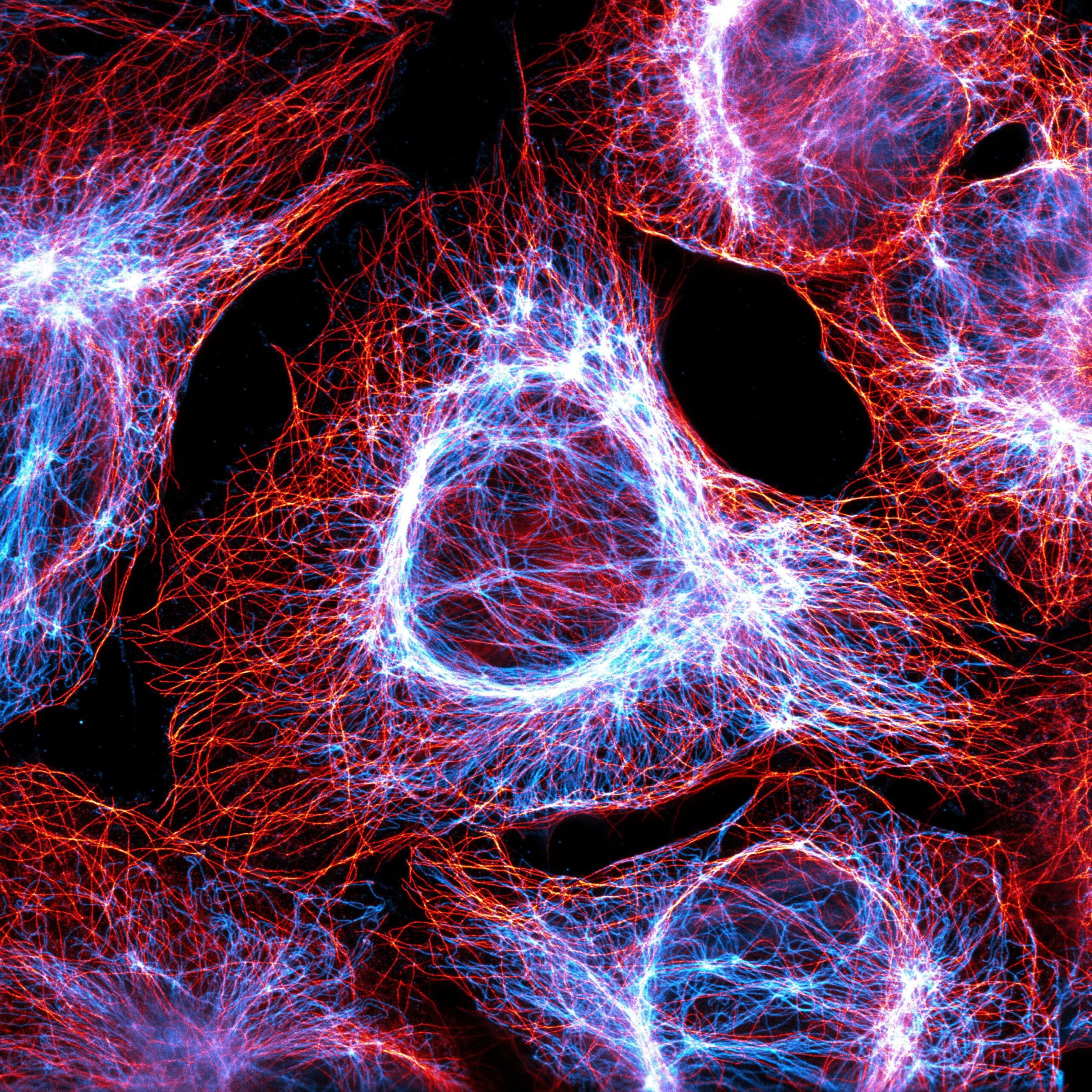
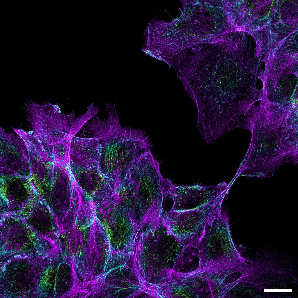
Dr. Till Stephan, Buchmann Institute for Molecular Life Sciences, Goethe University Frankfurt am Main
Intermediate filaments and microtubules visualized by super-resolution microscopy.

Anthoula Chatzimpinou, Weinhardt Lab
Microglia cell anatomy in 3D. Single microglia imaged with cryo soft-X ray tomography with visible keypoint organelles (nucleus, mitochondria, endosomes).
Matthias Rübsam, Niessen Lab, CECAD
Keratin10 filamentous network in the developing mouse epidermis. Fluorescence by endogenous expression of a Keratin10-NEON fusion (mouse generated by Hisham Bazzi). Color variation is due to height coding.
PD Dr. Christoph Gerhardt, UbiCilia Lab, Institute for Molecular Medicine (IMM), Health and Medical University (HMU) Potsdam
Cell Division with 3 Centrioles. Cell Type: immortalized kidney medullary (KM) cell line. Staining: detyrosinated α-Tubulin (green), gamma Tubulin (blue), Nphp1 (red).
Maik Bischoff and Sarah Clark, Peifer Lab, UNC Chapel Hill
Drosophila viridilis testis with musculature stained with phalloidin.
Lena Sarah Stanisławczyk (doctoral candidate), laboratory of Prof. Armin Huber at the Department of Biochemistry, University of Hohenheim.
Image depicts Drosophila photoreceptor cells, stained with α-F-Actin, α-Nuclei, α-TRPL, and α-Rab11-Myc.
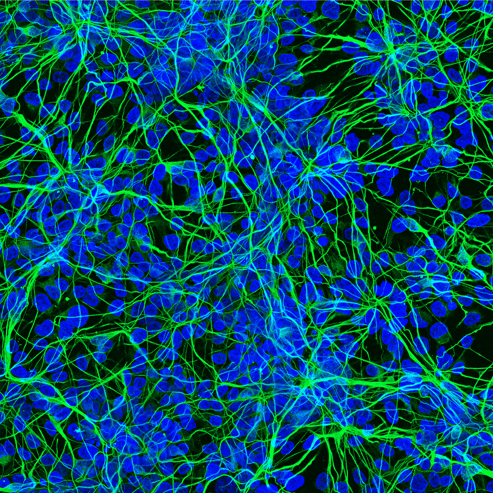
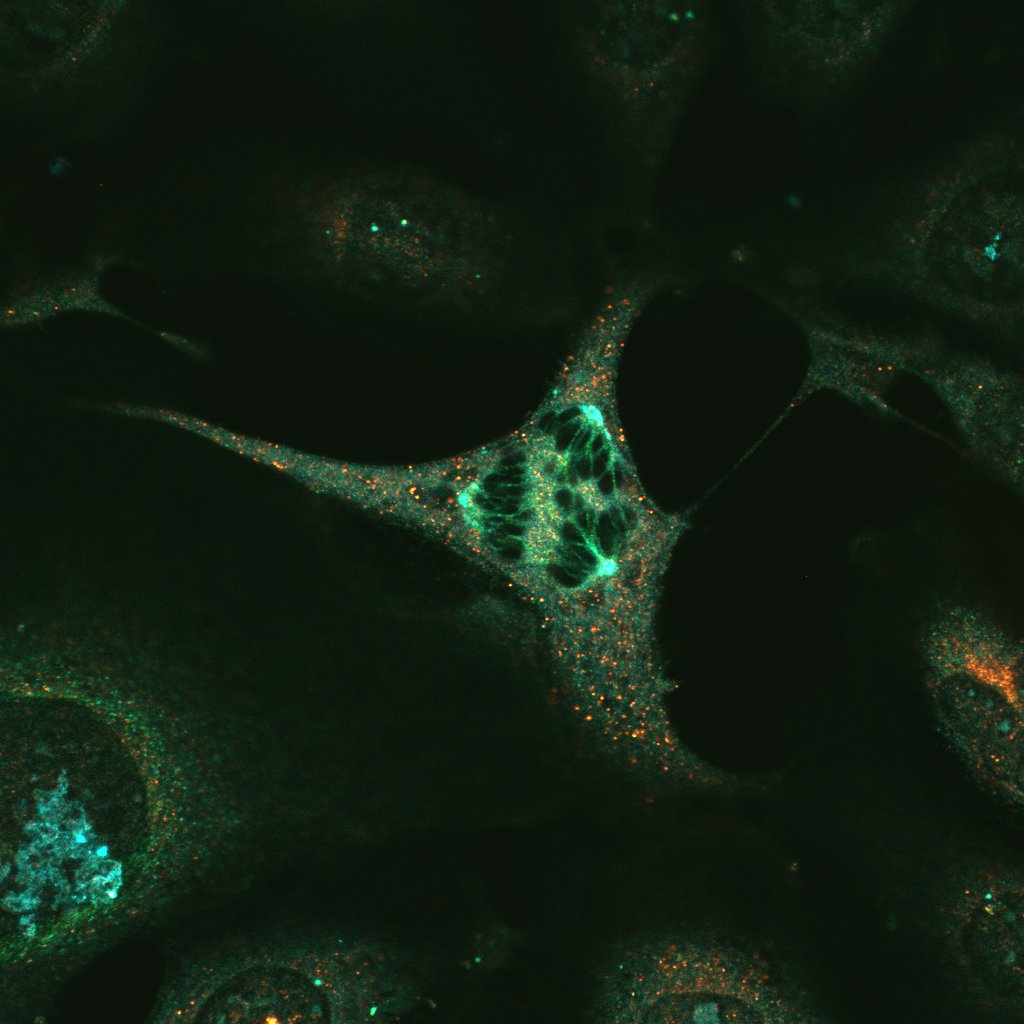
M. Sc. Maruthi Kumar Pabba (doctoral student), Cardoso lab
In vitro differentiation of human Induced pluripotent stem cells to mature neurons. The image shows nuclei labeled with DAPI (blue) and beta tubulin 3 (green) a neuronal marker.
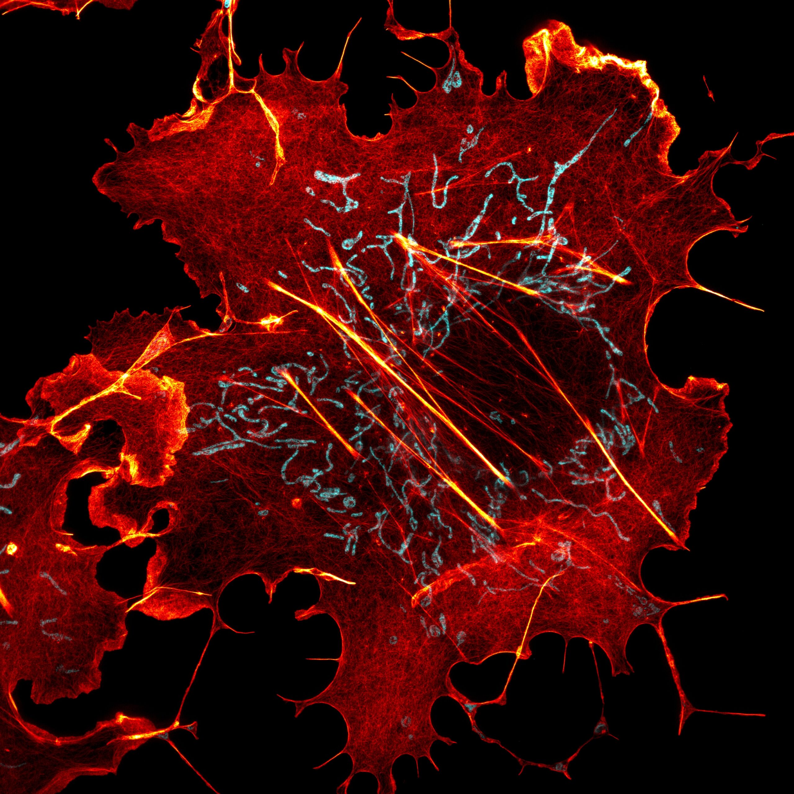
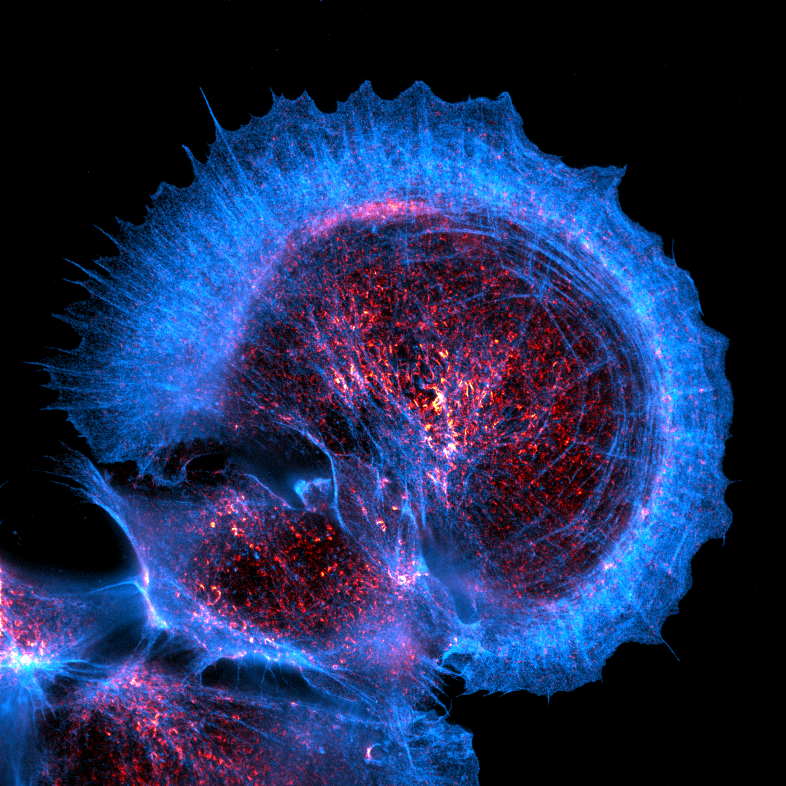
Dr. Till Stephan, Buchmann Institute for Molecular Life Sciences, Goethe University Frankfurt am Main
The actin cytoskeleton and mitochondria visualized by super-resolution microscopy.
Lena Strieker, PhD student Cavalcanti-Adam Lab/Cellular Biomechanics & Max Planck School Matter to Life
3D organization of actin in sphingolipid-deficient epithelial cells
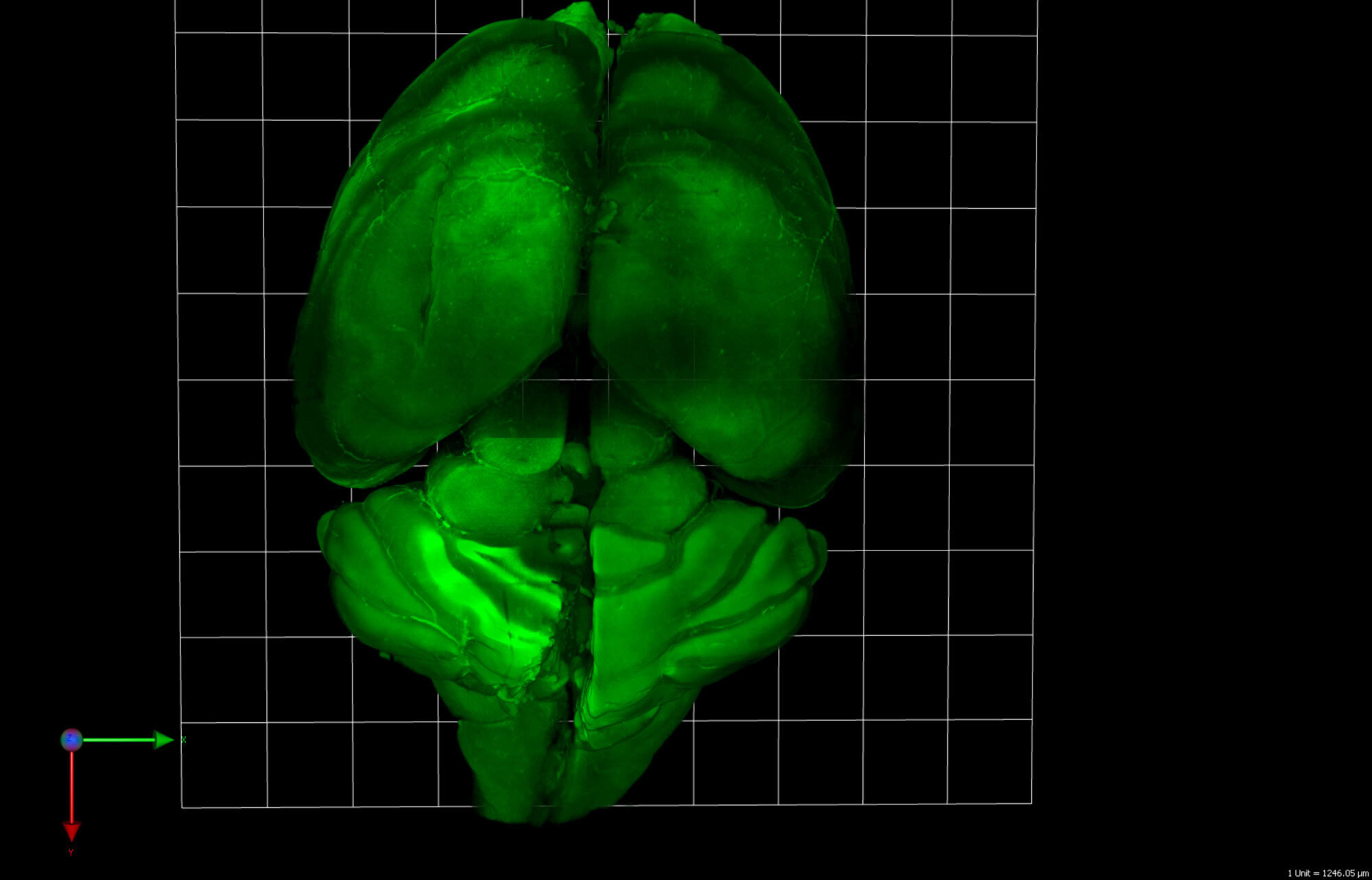
Ksenia Kolobynina, Vanessa Carlos, AG Cardoso, TU Darmstadt.
Mouse brain, 3D projection. Cleared and stained with methylene blue to label the nuclei. Light-sheet microscopy.
Eugen Kerkhoff
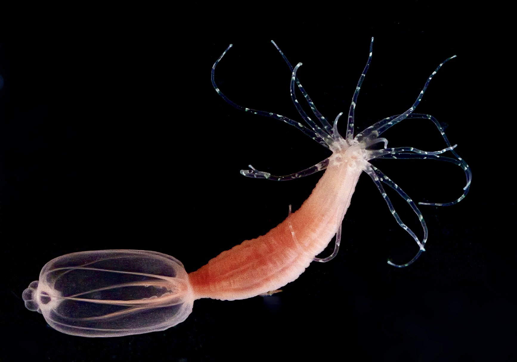
A picture of the sea anemone Nematostella vectensis is shown. Nematostella vectensis is an emerging animal model in the field of molecular cell biology. Tools to generate transgenic or knockout animals have been established and allow the functional characterisation of basic animal protein functions in a diversity of processes such as tissue regeneration or neuronal signalling. The picture has been taken with a Panasonic Lumix DC-S5M2X digital camera and a Sigma 70 mm F2.8 DG MACRO lens (aperture f13, exposure time 1/30 s). The picture was further processed in Adobe Photoshop.
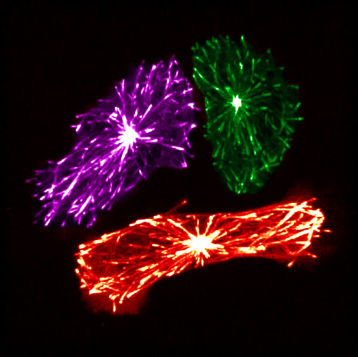
Germán Camargo Ortega, Cell Systems Dynamics, ETH Zürich
Microtubules in blood progenitors, all same but different.
Maik Bischoff, Peifer Lab, UNC Chapel Hill
Nuclei of pupal Drosophila pseudoobscura developing male gametes stained with Dapi.
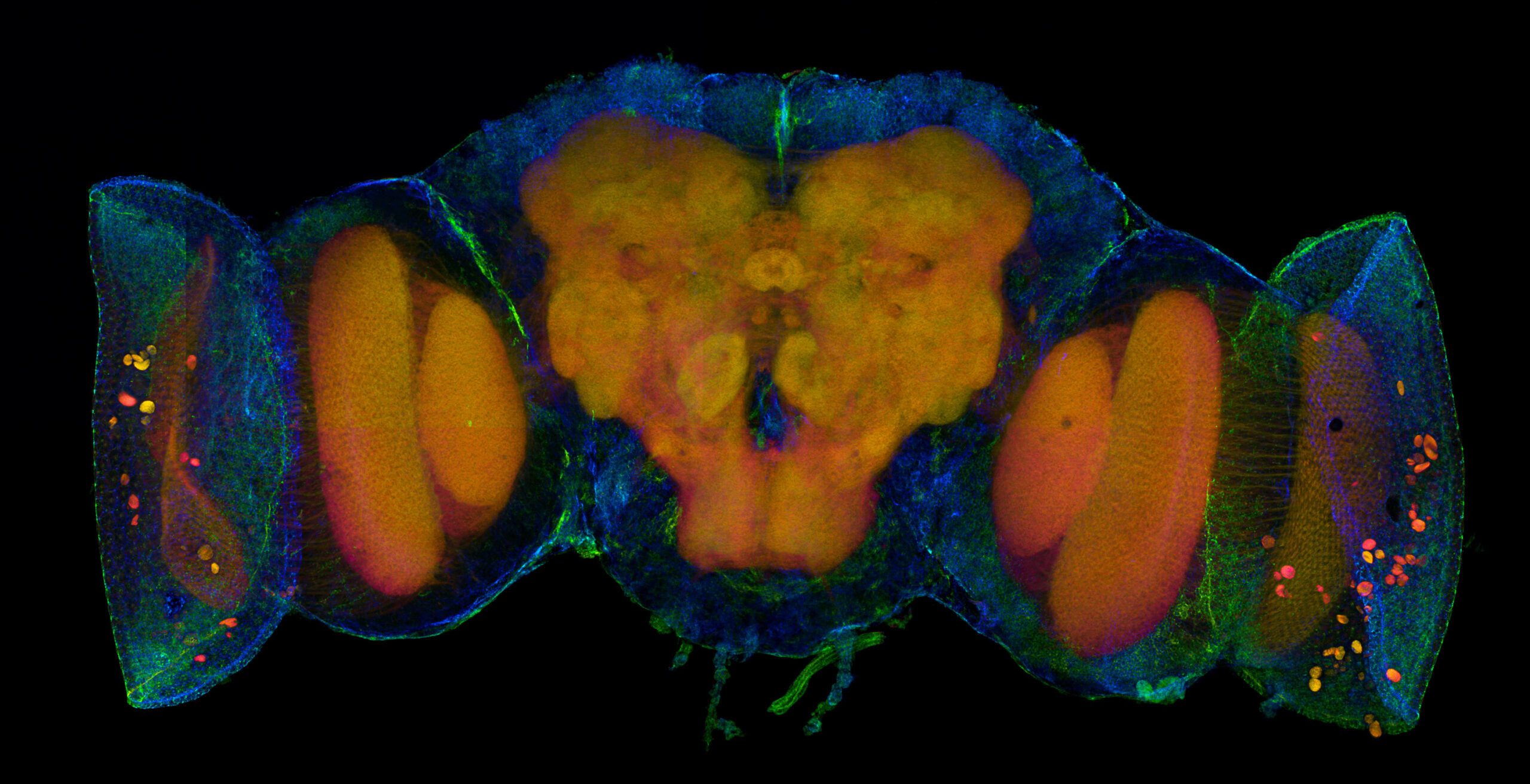
Maik Bischoff, Peifer Lab, UNC Chapel Hill
A pupal Drosophila Brain with Eye imaginal discs stained with Phalloidin and E-Cadherin.
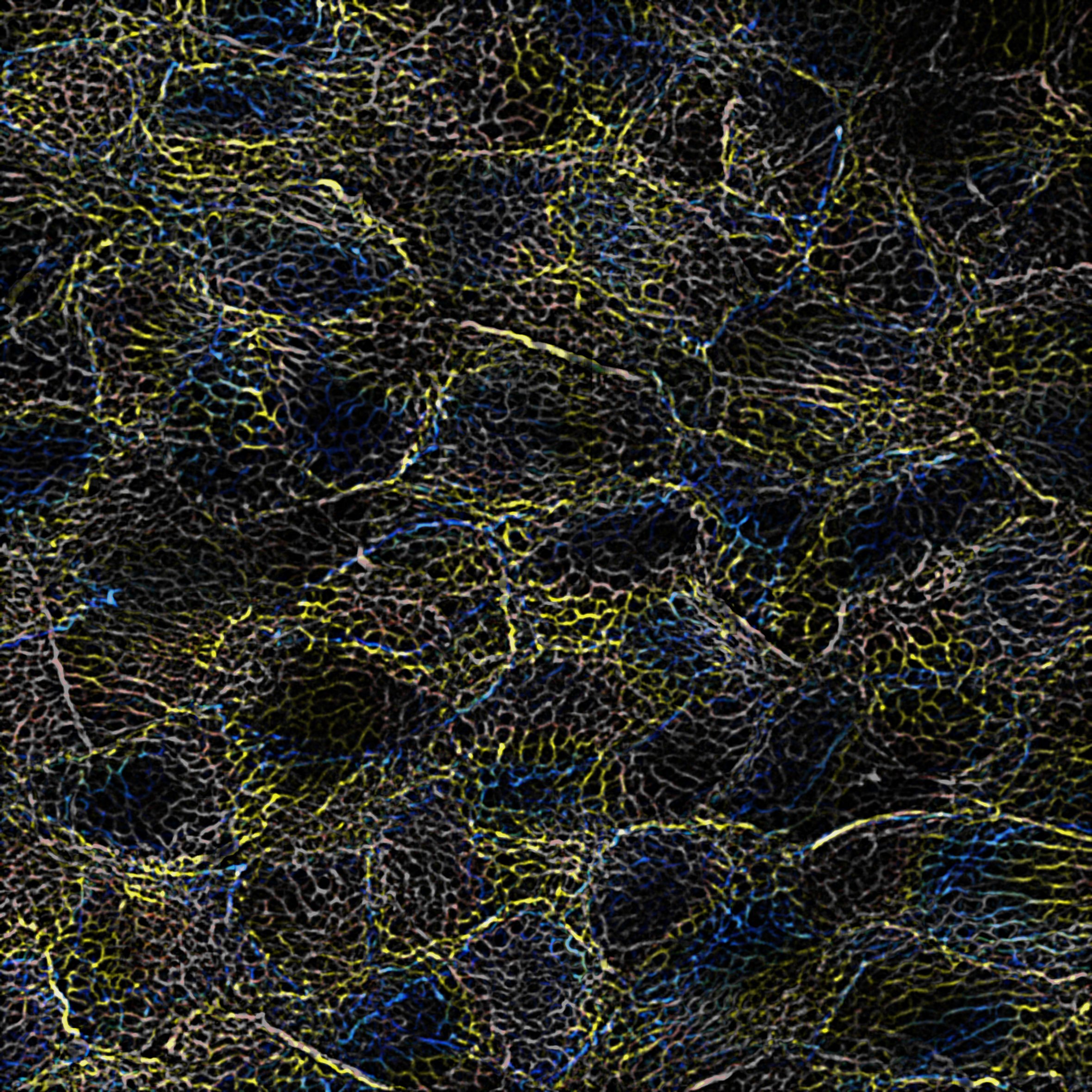
Max Gerlein and Paul Markus Müller, Ewers Lab, Freie Universität Berlin
STED image of NRK cells labeled for actin (blue) and septins (red).
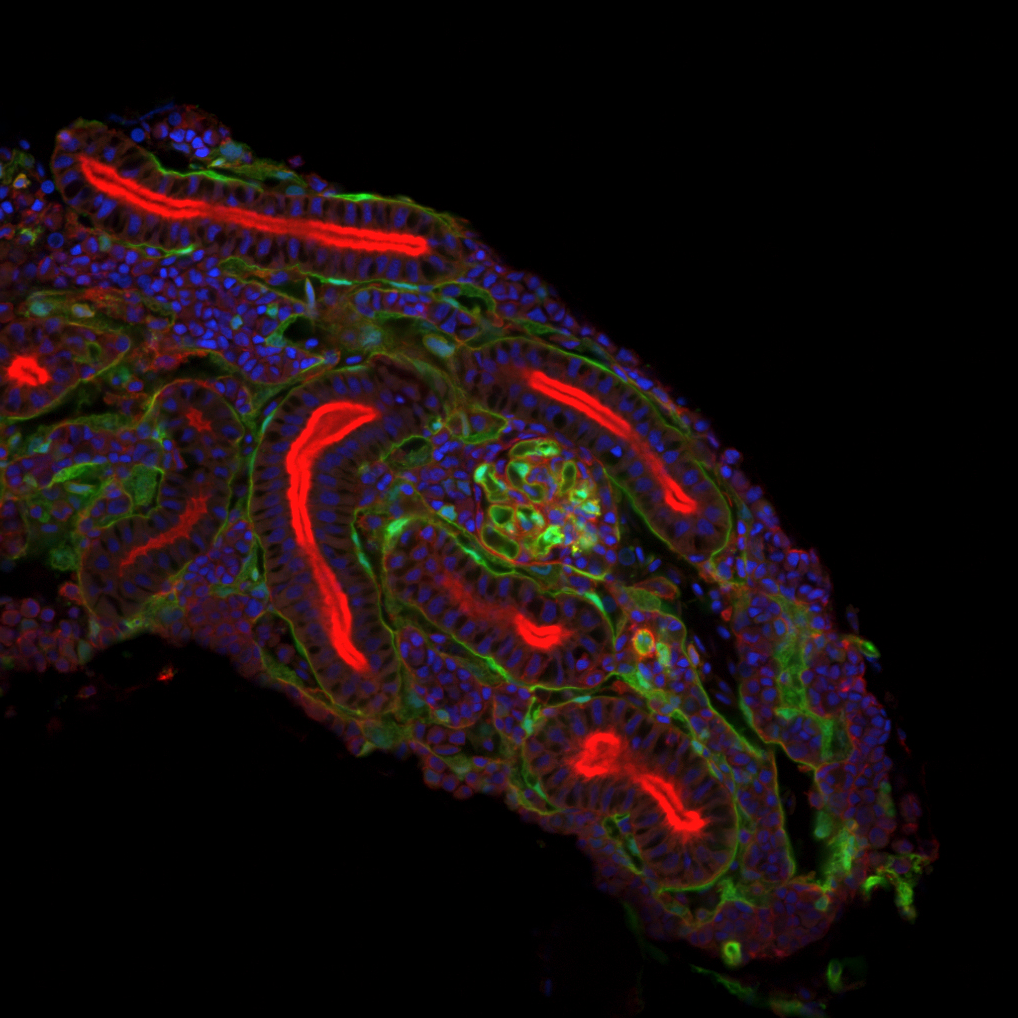
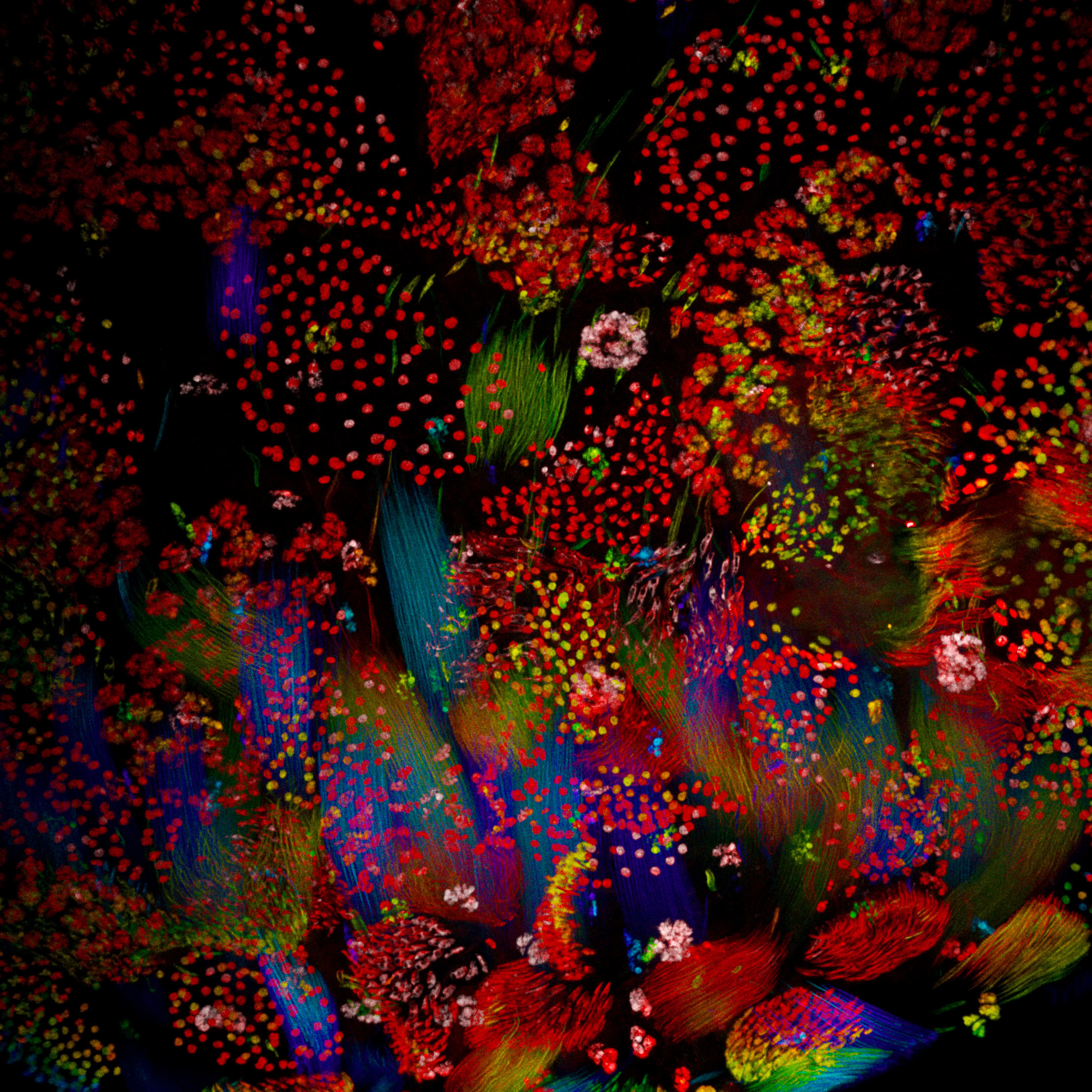
Mathieu Preussner, Lab: Virginie Lecaudey
Adult zebrafish mesonephros cryosection. Cryosection of the mesonephros from an adult fli1a:GFP Danio rerio, stained with Phalloidin (red) to label the tubular system and DAPI (blue) to stain nuclei. GFP is beautifully expressed in the capillary tuft of the glomeruli (middle).