Cell News 3
&
4/2016
17
MEETING REPORT
Meeting report
The 7th Annual Symposium Physics of Cancer took place from
October 4-6 in Leipzig, Germany, and was once again a vibrant
forum for the sharing of results and exchange of ideas at the
intersection of oncology and physical sciences. Approximately
100 participants took part in this year's meeting, half of which
were students, signaling a continued interest amongst the
current and up-and-coming generation of scientists. Due to
renovations at the traditional venue of the Biotechnological-
Biomedical Center (BBZ), the meeting was moved to the nearby
venue Haus des Buches. The organizing committee included
founding members Prof. Harald Herrmann and Prof. Josef
Käs, as well as new member Dr. David Smith, representing the
Fraunhofer Institute for Cell Therapy and Immunology. The
program included a total of 36 presentations, of which 28 were
from invited speakers and eight were selected from contributed
abstracts. Eight countries were represented, including Germa-
ny, Canada, France, USA, UK, South Korea, Israel and Sweden,
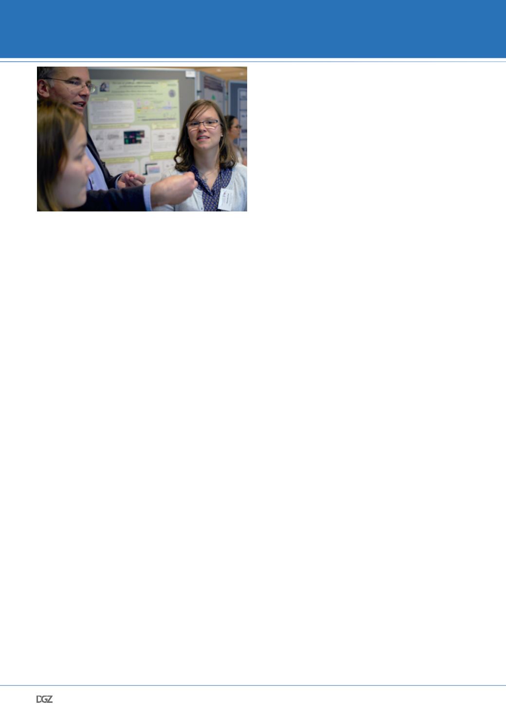
and included a total of nine female researchers. Throughout
the meeting, there were regular opportunities for participants
to talk in a comfortable, informal setting to share ideas or plan
future projects (Figure 1).
While it would be impossible to give each of the 36 presen-
tations the attention they deserve, several highlights of each
session are given in the following.
October 4th (Tuesday)
A brief welcome address was given by Prof. Josef Käs in the
early afternoon, greeting all participants to begin the con-
ference and setting the agenda for the following days. The
first subject area focused on the topic Functional Mechanics
of Cancer Cells, encompassing both sessions of the afternoon.
University of Pennsylvania professor Paul Janmey was the
opening speaker, and shared a breadth of results coming from
a wide survey of how different types of cancer cells - more
than 30 in all - react mechanically to substrate mechanics and
functionalization schemes. His group's recent work has shown
that substrates of hyaluronic acid (HA), a common extracellular
matrix polymer that is often upregulated in cancer, which are
functionalized with integrin ligands, can give rise to increased
invasive activity on the single-cell level. Specifically, the lo-
comotion, spreading and proliferation of most types of cancer
cells on even soft HA substrates is significantly increased when
compared to other types of substrates (e.g. polyacrylamide)
with similar stiffness. Later in the first session, Joachim Rädler
shared the recent work from his group at the Ludwig-Maximi-
lians-University in Munich, Germany, on the effect of micro-
patterned substrates on cell migration. Both microchannels
and patterns of adhesion-promoting molecules on substrates
were used to guide local cell interactions. Small collections
of cells confined in chambers tend to display a rotational be-
havior, dependent upon cell number, spatial arrangement and
internal polarity o the cells. Furthermore, "dumbbell" configu-
rations with multiple chambers connected by a channel - or in
some cases separated by a pseudo-barrier - also showed a va-
riety of novel cell migration behaviors, with hints of statistical
correlation between the relative occupancy of the chambers
versus the size, geometry and separation of the chambers.
In the second afternoon session, Allen Ehrlicher from McGill
University in Canada shared the results from his ongoing work
on determining how mutations in certain cytoskeleton-linked
molecules (specificallly alpha-actinin) can influence funda-
mental properties of cells such as internal dynamics, force
generation and locomotion. Most interestingly, the effects
of a specific mutation in actin crosslinker α-actinin 4 (ACTN4)
linked to kidney disease, was examined on the single-cell level.
The mutation is known to greatly increase the strength of
ACTN4 crosslinking, and was demonstrated to both slow cell
movement and internal cytoplasmic mobility, while increa-
sing their ability to exert forces on the surrounding substrate.
Afterwards, Rebecca Wells shared work indicating mechanical
commonalities between the development of fibrotic tissue
structures occurring during cirrhosis of the liver and the deve-
lopment of liver cancer. By carrying out rheological measure-
ments on liver tissue, a stiffening of cirrhotic tissue was found.
However, internal liver cancer tissue was found to display an
even higher stiffness, possibly due to the changes in the mic-
roenvironment such as increased proteoglycans and hyaluronic
acids. NIH researcher Kandice Tanner immediately continued
the theme of biological microenvironments, focusing much of
her presentation on the fabrication of 3D topographies through
the controlled self-assembly of magnetic particles functiona-
lized on their surfaces with extracellular matrix (ECM) prote-
ins. Closing the session and the first afternoon of talks, Lisa
McCawley from the Vanderbilt-Ingram Cancer Center shared
her group's technological advances in developing a microfluidic
platform where a simulated microenvironment, or "organ on a
chip" could be monitored and controlled in real-time, facilita-
ting the thorough testing of anti-cancer drugs on the single-
and multicellular level.


