Cell News 3
&
4/2016
14
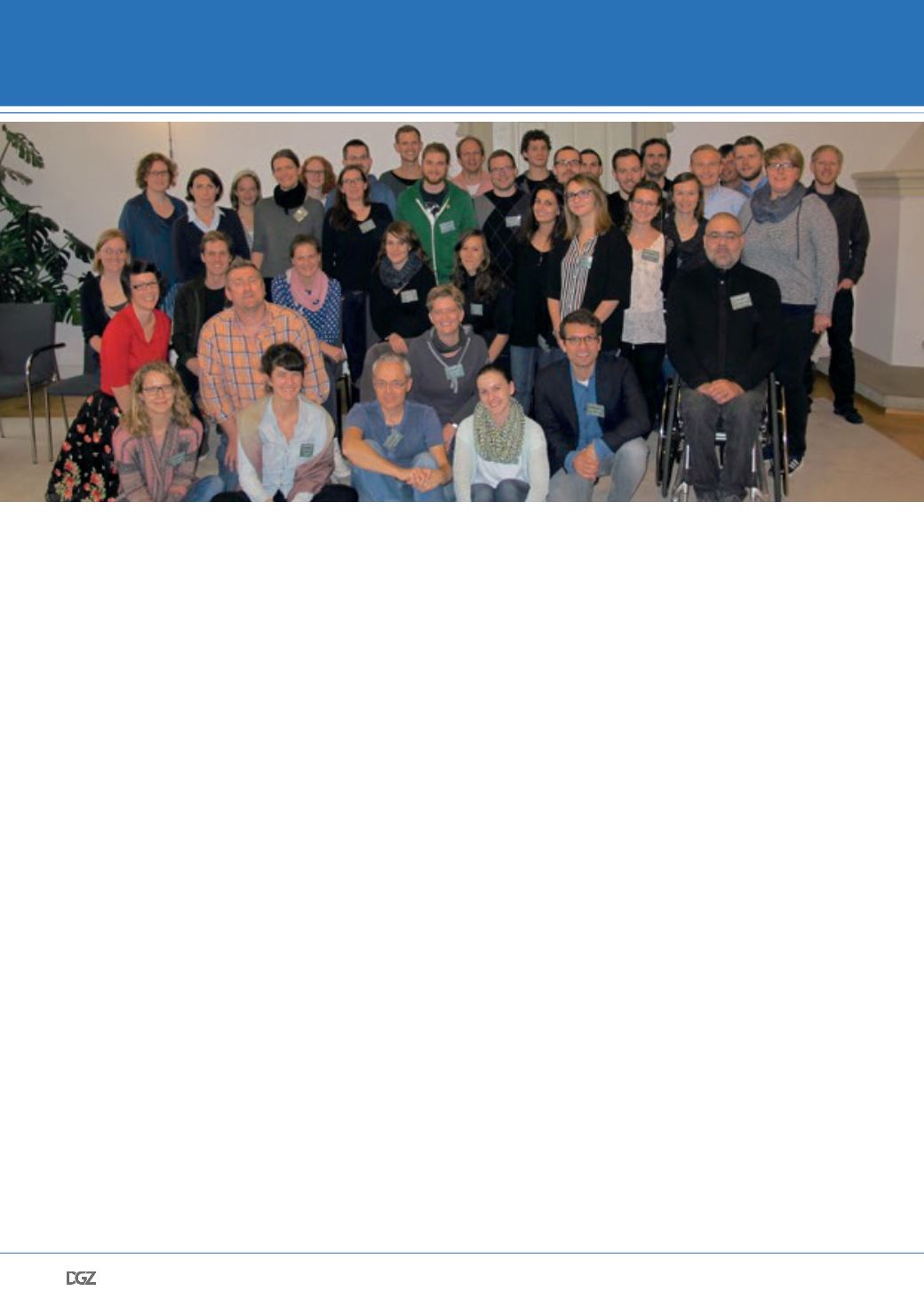
This year the workshop “Cell Biology of Viral Infections” celeb-
rated its 15th anniversary with the theme “Revolutionizing cell
biology tools for virology”. 43 people attended the workshop,
which included 25 students and 5 post-docs. The workshop has
continued its tradition as an open and friendly environment for
students to have fruitful discussion and get feedback and new
insights into their work. Throughout the years, we noticed that
students are more and more participative and are not afraid
of asking questions to each other and to the keynote speakers.
We are very proud that the workshop is successfully reaching
its objectives in making students more confident and opened
minded on various fields of research.
The workshop was kicked off by the keynote lecture from
Prof. Jochen Guck of the Technical University of Dresden. Prof.
Guck discussed his labs focus on understanding how mecha-
nical functions can be related to cellular changes. His lab has
developed cutting edge methods including microfluidic optical
stretchers and real-time deformability cytometry which allows
for high throughput measures of mechanical changes in cells.
These techniques are currently being used to analyze blood
samples to better predict infections, cancers, and abnormali-
ties.
The second keynote lecture was given by Prof. Khalid Salaita
from Emory University in Atlanta, GA, USA. Dr. Salaita’s work
focuses on understanding how mechanical forces influence
biological processes. As cellular forces can be so small, his lab
has developed key technologies to be able to measure forces
at the pN level. A key point for the students was the evolution
of these methods, as he showed three different generations of
force sensors and how his lab is constantly adapting and im-
proving its technologies. Prof. Salaita’s lab is now using these
force sensors to ask questions about “mechano-pharmacology”
where they are able to understand how lung cells response to
asthma medications. They hope to apply these sensors to many
other medical fields and science disciplines in the near future.
Prof. Matthias Gunzer from the University of Essen delivered
the third keynote address. His lab has pioneered imaging
methods to enable the visualization of whole tissues from ani-
mals. They are inspired by the idea of “can you count this”. This
has pushed the Gunzer lab to develop computation methods to
be able to quantify important features in a tissue. Using this
technology, they can understand how the brain changes after
a stroke and which cell populations are most affected. This
understanding has then helped them evaluate medications that
if given soon enough after onset, can decrease the harmful
effects of the stroke. In addition, the Gunzer lab is developing
methods to track cell migration of neutrophils. These efforts
have allowed them to detect onset of disease prior to onset of
symptoms. Additionally, these methods have allowed them to
evaluate patients with neutrophil disorders and they are now
able to determine which therapies will restore the neutrophil
populations to normal levels and behaviors.
The final keynote lecture was given by Dr. Kem Sochaki. Dr. So-
chaki is a staff scientist at the National Institute of Health in
Bethesda, USA. As her training was in microscopy methods, she
related to the students how important it is to look at an image
15TH WORKSHOP “CELL BIOLOGY OF VIRAL
INFECTIONS” OF THE SOCIETY FOR VIROLOGY (GFV)
MEETING REPORT


