aspects of the field had to be neglected given the space con-
straints. We asked the authors not only to cover recent
findings, but also to provide their views on the issues at
stake and emphasize important questions for the future. We
articulated the 16 contributions into four thematic groups: cen-
trosomes in history and evolution, centrosome assembly and
structure, the functions of centrosomes, as well as centrosomes
in development and diseases. In addition, the present piece
serves as a preface, whereas an exceptional account by
Ulrich Scheer on Boveri’s years in Wu¨ rzburg, with newly dis-
covered plates of his work, follows as a prologue and as a
reminder of the origin of a field started over a century ago [5].
In order to have a Theme Issue that is representative of the
main advances and concepts in the field, each contribution has
been reviewed by two or three experts who could have written
just as well on the particular topic they have been asked to
review. Those reviewers who were willing to have their
names disclosed are listed at the end of this preface. In this
manner, in addition to the two undersigned who acted as
joint editors for every chapter, the contents of this Theme
Issue reflect the direct or indirect input of some 50 leading
scientists in the field, whom we wish to thank wholeheartedly
for their important contributions.
3. The main characters
In this preface, we attempt to set the stage for the Theme Issue,
while avoiding redundancies with the individual contributions
to the extent possible, such that the reader is invited to consult
the respective papers for further information and references.
As in all fields, but especially in ones that span more than a
century, during which conceptual frameworks and experi-
mental approaches have changed substantially, there is a need
to ensure some shared basic terminology to facilitate communi-
cation between members of the community and accelerate entry
into the field for newcomers. For instance, until when should a
procentriole be referred as such before being called a centriole in
the canonical centrosome duplication cycle? Hereafter, we use
the term procentriole to refer to a centriolar cylinder from the
moment it is discernible next to the proximal end of a parental
centriole, approximately at the G1/S transition, until mitosis of
that cell cycle (figure 1
a
). During this time interval, a procen-
triole and the parental centriole next to which it emerges are
referred to collectively as a diplosome. After disengagement of
the procentriole from the parental centriole during mitosis and
subsequent entry into G1, the younger structure, which used
to be the procentriole, is now referred to as the daughter
intercentriolar linker
(
a
)
(
b
)
A B C
A B
sub-distal
appendage
distal
appendage
1
2
3
cartwheel
hub
cartwheel
spoke
1
2
3
satellites
procentriole
cartwheel
sub-distal
appendages
distal
appendages
PCM
daughter
centriole
mother
centriole
procentriole
B
A
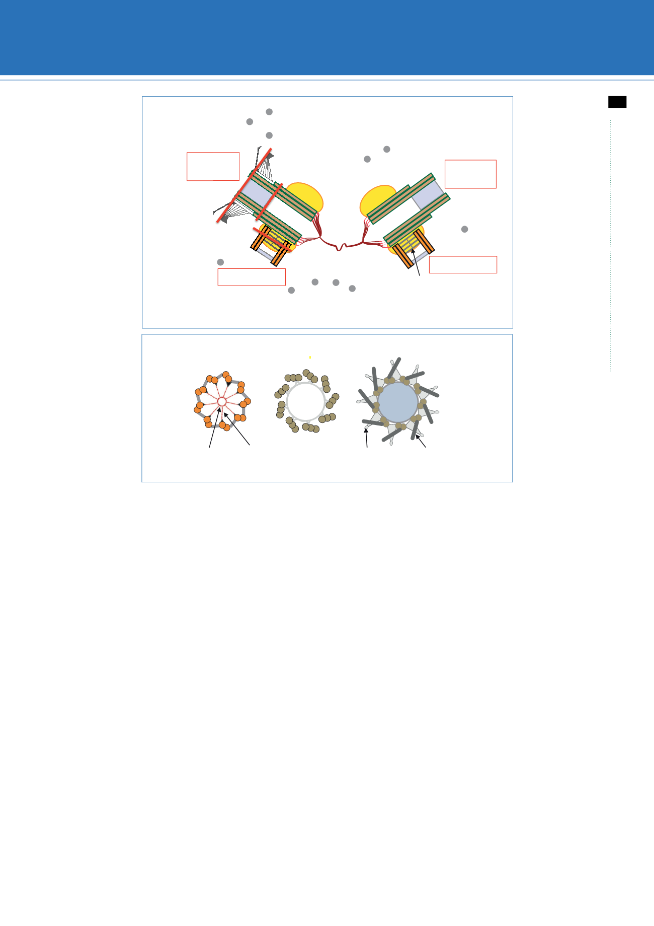
Figure 1.
Centrosomes in human cells. (
a
) Representation of a pair of centrosomes in human cells viewed from the side during the S phase of the cell cycle. The
lines designated 1, 2 and 3 indicate the positions corresponding to the cross sections shown in (
b
). The parental centrioles are approximately 450 nm long and
approximately 250 nm in outer diameter. The grey region in the distal part represents the filled lumen in the region where centrin concentrates [6]. Note that for
simplicity the cartwheel is represented with only four slices and that it is present only in the procentriole in human cells. Similarly for simplicity, the PCM/
centrosomal matrix is represented solely around the proximal region, even though it is also present to a lesser extent around the more distal segments.
(
b
) Corresponding cross sections, viewed from the proximal end, in regions 1 (proximal part of the centriole, with cartwheel highlighted and triplet microtubules
denoted A, B, C), 2 (central part of the mother centriole, also with triplet microtubules) and 3 (distal part of the mother centriole, with double microtubules denoted
A, B). (Online version in colour.)
rstb.royalsocietypublishing.org
Phil. Trans. R. Soc. B
369
: 20130452
2
on October 7, 2014
rstb.royalsocietypublishing.org
Downloaded from
Book Recommendation


