Cell News 02/2017
18
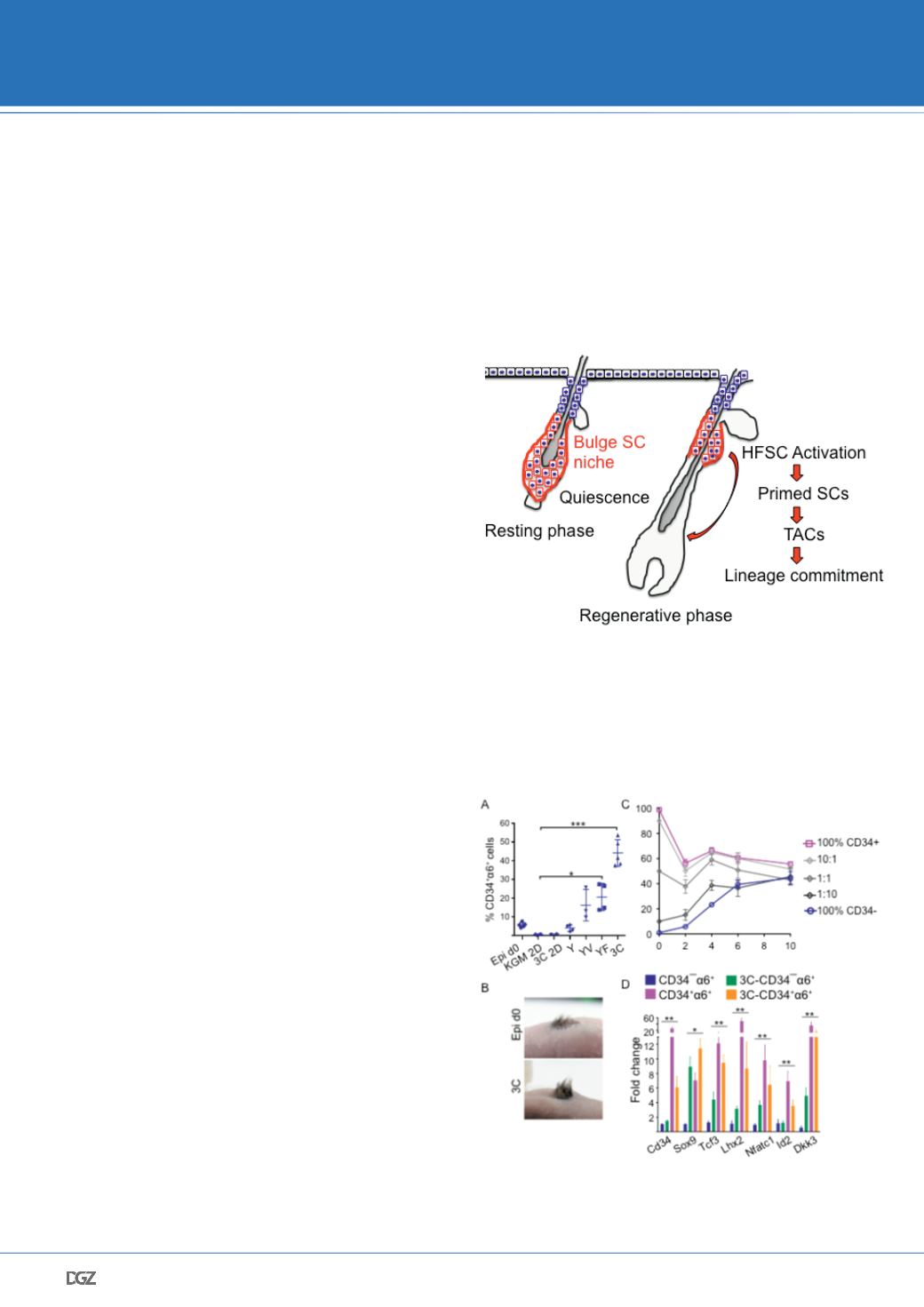
Quiescent HFSCs reside in the bulge niche within hair follicles
where they are activated in a two-step process to proliferate, to
leave the niche, and to differentiate in order to regenerate the
hair follicle.
Mouse epidermis (Epi d0) cultured under various conditions
show that 3C conditions (3-dimentional Matrigel with keratino-
cyte growth medium (KGM) supplemented with VEGF (V), bFGF
(F), Y2732 (Y)) enrich for HFSCs (CD34+
α
6+), whereas 3C medi-
um in 2-dimentional culture (3C 2D) does not support growth.
(B) Cultured HFSCs retain their multipotency as shown by trans-
plantation assays into nude mice. (C) HFSCs (CD34+) and non-
HFSCs (CD34-) self-evolve into a 50:50 population equilibrium
over time. The equilibrium can be achieved from either pure
HFSCs or non-HFSCs, or any mixed ratio of the two populations
(D) 3C-HFSCs (in orange) express key lineage identity transcription
factors and closely resemble
bona fide
CD34+
α
6+ HFSCs directly
isolated from mouse epidermis (in pink), whereas 3C progenitors
(CD34-
α
6+; in green) express intermediate levels of stem cell
genes, when compared to freshly purified
in vivo
progenitors (in
blue). Modified from Chacon-Martinez et al., 2016
BINDER INNOVATION PRIZE 2017
Figure. 1 Schematic illustration of HFSC regulation in their bulge niche
Figure 2: HFSC cultures reveal dynamic bi-directional plasticity between SCs and progenitors. (A)


