Cell News 4/2014
18
Rac-GEF Tiam1, thereby coupling Rac activation to Par polarity
signaling. Cross-talk between polarity complexes and Rho GTPa-
ses is necessary to accomplish the cytoskeletal changes required
for polarization of various cell types (Iden and Collard, 2008).
Epithelial polarity: an overview
Epithelial sheets cover organs and body cavities, such as the
lung, gut or skin. Epithelia form important dynamic barriers bet-
ween the external environment and the interior of an organism,
and impart mechanical protection or mediate secretion, absorp-
tion and sensory functions. Epithelial integrity and homeosta-
sis are of central importance to survival, and mechanisms have
evolved to ensure these processes are maintained during growth
and upon damage. Different types of epithelial junctions that
connect the cells to the underlying tissue or to their neighbors
(fig. 1A) mediate adhesion or barrier functions, and sense and
respond to mechanical forces and cell density. Epithelial junc-
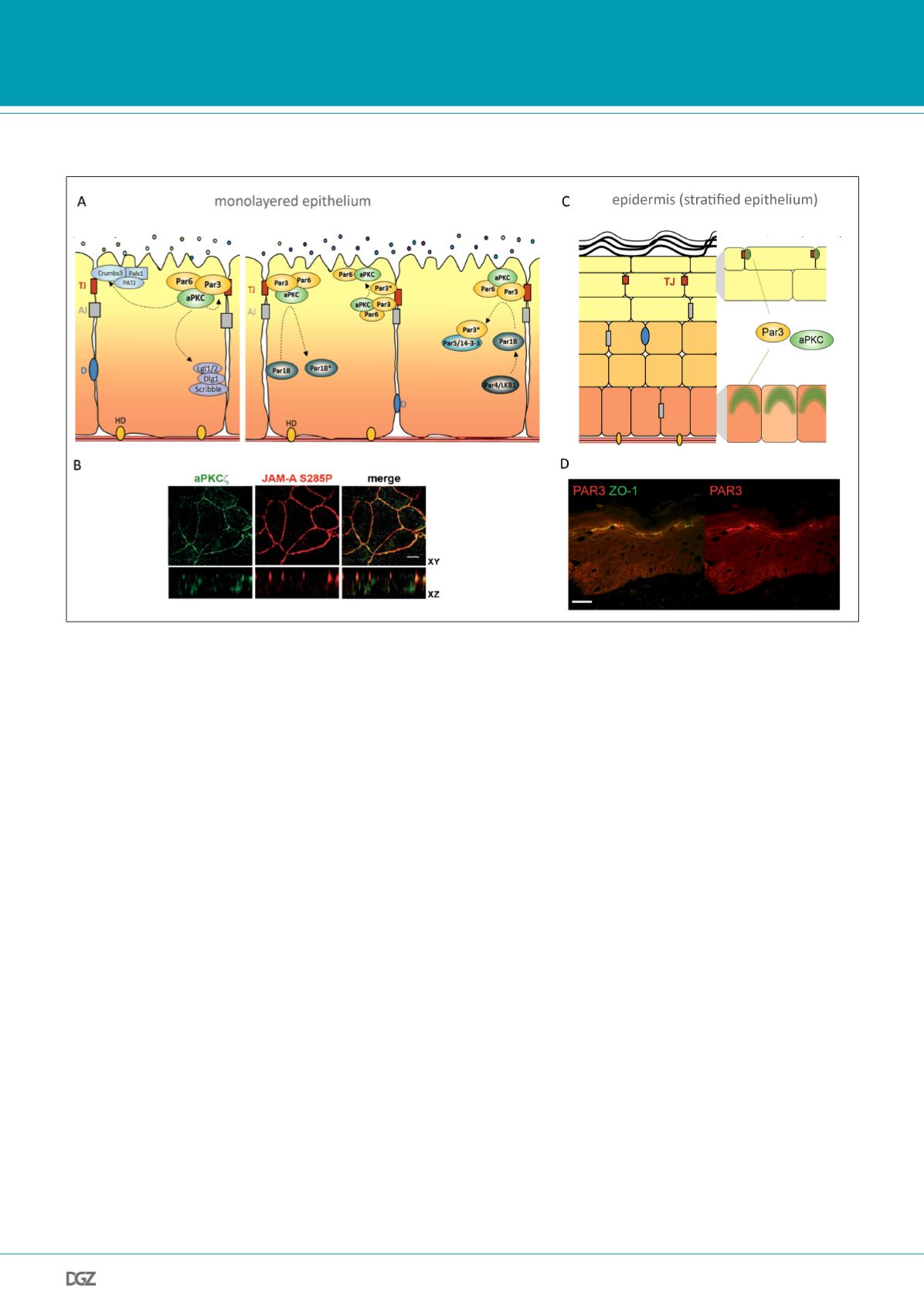
Figure 1: Overview of mammalian polarity proteins in simple epithelia and stratified epidermis.
A, At the example of cultured mammalian monolayered epithelial cells, the subcellular localization and mutual exclusion of polarity proteins is illustrated.
Left panel: Localization of the three “classical” polarity complexes, Par3-aPKC-Par6, Crumbs3-Pals1-PATJ, and Scribble-Dlg1-Lgl1/2 in a mammalian simple
epithelium. Right panel: Par3 and Par6 are scaffold proteins that interact with the Ser/Thr kinase atypical PKC (aPKC). The Par3 complex localizes to tight
junctions (TJs) and is required for proper TJ formation of simple epithelia. During apical domain assembly, Cdc42-mediated activation of aPKC results in
phosphorylation of Par3 and dissociation of aPKC/Par6 to the apical plasma membrane. Other mammalian Par proteins include Par1/MARK, Par4/LKB1 and
Par5/14-3-3. The Ser/Thr kinase Par1B is able to phosphorylate Par3, thereby regulating basolateral exclusion of Par3, which is subsequently bound by
Par5/14-3-3 through phospho-serine recognition motifs in Par5. aPKC-mediated Par1B phosphorylation mediates the dissociation of Par1B away from the
cortex. The tumor suppressor protein Par4/LKB1, a Ser/Thr kinase, phosphorylates a variety of downstream targets including Par1B and the tumor suppres-
sor protein kinase AMPK (not shown), thereby controlling a range of cellular effects including intercellular adhesion, polarization, and energy metabolism.
B, Immunofluorescence micrograph of apical-junctional localization of aPKC and the phosphorylated form of the transmembrane protein JAM-A in pola-
rized murine mammary epithelial cells (MTD-1A). Scale bar: 5um (modified from Iden et al., 2012a).
C, Schematic representation of human stratified epidermis and the context-dependent localization of Par3 and aPKC. In presence of TJs, which are formed
in the outer viable layer of the epidermis, the stratum granulosum, Par3 and aPKC localize to these junctions. In contrast, in basal layer cells that lack TJs,
Par3 and aPKC show more diffuse cytoplasmic staining with some enrichment in the apical cortex. D, Immunofluorescence micrograph of Par3 and ZO-1, a
peripheral TJ protein, in human epidermis. Scale bar: 10um (modified from Iden et al., 2012b). TJ, tight junction; AJ, adherens junctions;
D, desmosomes; HD, hemi-desmosomes; *: phosphorylated protein.
tions are also important sites of signal transduction. Through
robust cell–cell and cell–matrix adhesions epithelial cells are
able to form diverse organized structures, including tubes, al-
veoli and stratified sheets. Epithelial intercellular junctions are
hallmarks of epithelial polarity, and formation and maintenance
of junctions is intimately linked to cell polarity signaling.
The epidermis: a self-renewing stratified epithelium
Mammalian skin protects the organism from dehydration and
provides a barrier against harmful influences, such as UV, tem-
perature and microbes. The epidermis forms the outermost sheet
of the skin and consists of the interfollicular epidermis (IFE) and
epidermal appendages, such as hair follicles, sebaceous and
sweat glands. Epidermal keratinocytes balance life-long self-
renewal in the proliferating basal layer with a tightly regulated
terminal differentiation program that is necessary to form the
stratum corneum, a dead, cornified and water-impermeable cell
Research news


