Cell News 3/2013
15
RESEARCH NEWS
Random pairing
In consequence, we will now turn to the question of how the
precursor cells are arranged immediately before fusion. Yield
is one important parameter for assessing the applicability of a
method for a given problem at hand, throughput is another one.
Macrosystems for cell fusion almost exclusively use random pai-
ring of cells, while the advent of reproducible and affordable
microfluidic systems allowed for progressing towards selective
pairing. Some microsystems use random pairing nonetheless for
reasons of ease and since random pairing is clearly correlated
with a high throughput. Such systems do not require extra pre-
paration time on cell pairing. Both cell types are simply mixed
together, then positioned and finally fused.
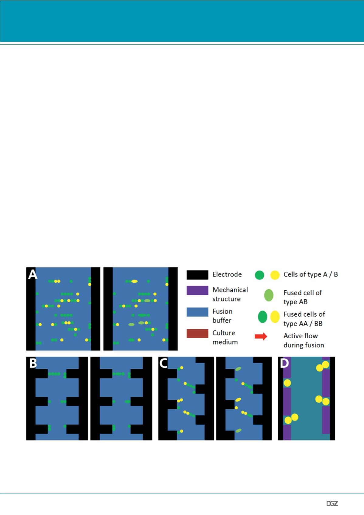
The simplest way of electrofusion are flat electrodes at the edges
of microchannels (see Figure 1A) as introduced by the group of
Ulrich Zimmermann (2). They mixed B cells and myeloma cells at
a ratio of 5 : 1, transferred them into fusion buffer, flushed them
into the fusion chamber and then aligned them dielectropho-
retically into a pearl chain configuration using an ac field. The
pairing consequentially is random. Subsequently, one or more dc
pulses of microsecond duration and high electric field strength,
i. e. 3.5 kV cm
-1
, were applied. The ac field remained on for some
seconds after the pulse. This leads to randomly located fusion of
two or sometimes more cells. After that, the cells were flushed
out, transferred into medium and pipetted into well plates for
HAT selection. Obviating any trapping or positioning, the pairing
and following fusion are necessarily random. Moreover, since
there is no successive sorting step, the fusion yield is low. On the
other hand, the throughput is very high. This also holds for the
economic success of the approach; and it still constitutes the
most widely adopted standard solution in hybridoma formation.
This system was improved by Tresset (3) and Hu (4) who desi-
gned new electrode forms for a more precise positioning and,
therefore, higher control over the fusion process (see Figure 1B).
While cell pairing was still random in this case, a major advan-
cement was made by using strongly non-uniform electric fields
which were produced by microelectrodes structured in a ridge-
like manner. By that, fusion was mainly induced in those cell
pairs that were located directly at the electrodes. With human
HEK-293 cells or protoplasts, fusion yields of (42 ± 2)% were ob-
tained and with HEK-293 cells and murine NIH/3T3 fibroblasts
of (60 ± 30)% (5-6). A further step ahead in the direction of
this structural concept were small silicon microcavities for cell
capturing (7), giving efficiencies of (70 ± 10)% with NIH/3T3
cells and myoblasts. Additional progress was made by modifica-
tions of the fluidic periphery, viz. through replacing the costly
syringe pumps by capitalizing on fluid motion by surface tension
Figure 1. Electrofusion after random cell pairing:
(a) Design scheme of the cell alignment in microchambers with flat electrodes before and after the pulse application (9). Pairing as well as fusion occur
randomly.
(b-c) Shaped microelectrodes improve the fusion efficiency due to creating non-uniform electric fields (8, 6). Fusion occurs preferentially of pairs located
near the electrode tips.
(d) Silicon microcavities for creating inhomogeneous electric fields (7). This additionally improves the capturing of groups consisting of only two cells.


