Cell News 3/2013
22
large extracellular domains and are directly or indirectly con-
nected to the cell wall (CW). This complex cell-spanning polymer
network is only slowly remodeled outside of active growth re-
gions and might represent a static scaffold. We have now started
to determine how CW synthesis and CW associated components
are distributed, and whether they influence distribution and mo-
bility of PM proteins. In plants, the influence of the cell wall
on cell cortex organization and lateral mobility of PM proteins
has already been documented
12
. Even proteins with small extra-
cellular domains are immobilized through their association with
the cell wall. In addition, the cell wall biosynthetic machinery
in rod-shaped bacteria, has been shown to actively move pro-
tein complexes in the PM and associated actin-like cytoskeletal
structures
17,27,28
.
A systems approach to cell cortex organization
To establish the relative importance and interplay between the
different mechanisms for cell cortex organization it will be
necessary to comprehensively analyze the mechanisms for la-
teral membrane segregation within a single system. The yeast
cell cortex constitutes a perfect model organism for this task.
Observation of PM domains in yeast is facilitated by their large
size and temporal stability. In addition, we have already estab-
lished powerful microscopy techniques to visualize the yeast cell
cortex with high sensitivity and contrast
16
. Finally, yeast cells
offer a vast array of resources for the systematic dissection of
molecular pathways.
One research field, where a quantitative understanding of the
factors driving cortex organization has proven invaluable, is cell
polarity. During polarity establishment in yeast budding we have
shown that the initial formation of a polarized site by the pola-
rity regulator Cdc42 is determined by a tightly regulated inter-
play between lateral membrane mobility, membrane recycling
and scaffolding
29-31
.
We are currently in the process of establishing multimodal high
throughput screens based on TIRFM to investigate factors af-
fecting PM organization. We will use automated microscopy to
image a large number of PM patterns that cover the whole diver-
sity of membrane environments and then systematically expose
these patterns to genetic, chemical or environmental perturba-
tions. These perturbations will be directed towards cellular lipid
composition, membrane trafficking, cytoskeletal dynamics and
cell wall biosynthesis. Additional factors that might influence
cortex organization are the recently described ER-PM contact
sites
32,33
. Importantly, the use of a genetically amenable in vivo
systems allows us to easily test the functional relevance of any
effect on PM patterns that we identify in our screen. For exam-
ple, it will be exciting to find in what manner nutrient uptake
and metabolic activities are affected by lateral partitioning of
the respective PM transporters and channels – with potential
impact on the large field of yeast biotechnology.
Ultimately, our work is expected to provide new insights into the
many cellular processes intimately linked to the plasma mem-
brane, such as signal transduction, protein turnover or inter cel-
lular communication. Even efforts in synthetic biology to create
minimal units of life will require a detailed understanding of the
self organization properties of biological membranes.
Acknowledgements
I would like to thank all the present and past members of the lab and my colleagues at the
University of Münster and the MPI of Biochemistry. Work in my lab has been funded by the
Max Planck Society, the Human Frontiers Science Program and the DFG (SFB863, SPP1464,
CiM cluster of excellence).
References
1 Arinaminpathy, Y., Khurana, E., Engelman, D. M. & Gerstein, M. B. Computational analy-
sis of membrane proteins: the largest class of drug targets. Drug Discov Today 14, 1130-1135,
doi:10.1016/j.drudis.2009.08.006 (2009).
2 Fiegl, M. et al. Physiological hypoxia promotes lipid raft and PI3K-dependent activation
of MAPK 42/44 in leukemia cells. Leukemia 24, 1364-1367, doi:10.1038/leu.2010.94 (2010).
3 Gajate, C. & Mollinedo, F. Lipid rafts and Fas/CD95 signaling in cancer chemotherapy.
Recent Pat Anticancer Drug Discov 6, 274-283, doi:10.2174/157489211796957766 (2011).
4 Mueller, N. S., Wedlich-Soldner, R. & Spira, F. From mosaic to patchwork: matching lipids
and proteins in membrane organization. Mol Membr Biol 29, 186-196, doi:10.3109/096876
88.2012.687461 (2012).
5 Anderson, R. G. & Jacobson, K. A role for lipid shells in targeting proteins to caveo-
lae, rafts, and other lipid domains. Science 296, 1821-1825, doi:10.1126/science.1068886
(2002).
6 Bagatolli, L. A., Ipsen, J. H., Simonsen, A. C. & Mouritsen, O. G. An outlook on organiza-
tion of lipids in membranes: Searching for a realistic connection with the organization of
biological membranes. Prog Lipid Res (2010).
7 Lingwood, D., Kaiser, H. J., Levental, I. & Simons, K. Lipid rafts as functional heterogenei-
ty in cell membranes. Biochem Soc Trans 37, 955-960, doi:10.1042/BST0370955 (2009).
8 Shaikh, S. R. & Edidin, M. A. Membranes are not just rafts. Chem Phys Lipids 144, 1-3,
doi:10.1016/j.chemphyslip.2006.06.017 (2006).
9 Douglass, A. D. & Vale, R. D. Single-molecule microscopy reveals plasma membrane
microdomains created by protein-protein networks that exclude or trap signaling molecules
in T cells. Cell 121, 937-950 (2005).
10 Charrin, S. et al. Lateral organization of membrane proteins: tetraspanins spin their web.
Biochem J 420, 133-154, doi:10.1042/BJ20082422 (2009).
11 Sackmann, E., Lipowsky, R. & Sackmann, E. in Handbook of Biological Physics Vol. Volu-
me 1, Part 1 1-63 (North-Holland, 1995).
12 Martiniere, A. et al. Cell wall constrains lateral diffusion of plant plasma-membrane
proteins. Proc Natl Acad Sci U S A 109, 12805-12810, doi:10.1073/pnas.1202040109 (2012).
13 Kusumi, A., Sako, Y. & Yamamoto, M. Confined lateral diffusion of membrane receptors
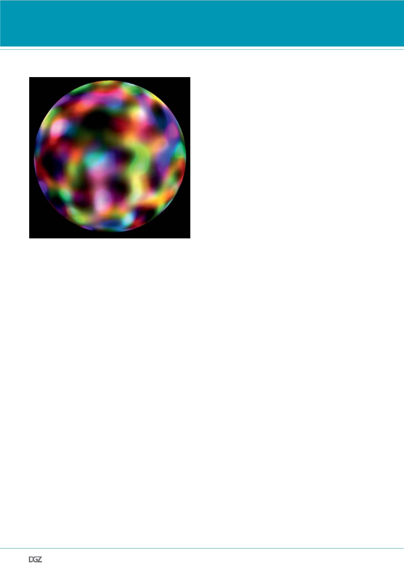
Figure 4.
Artistic representation of the yeast PM as molecular patchwork. Six ran-
domly overlapping domains from different original cells were labeled with
different colors and overlaid in Photoshop.
RESEARCH NEWS


