cell news 2/2013
8
characterized by cortical fows and circumferential constriction;
both occurring with velocities on the order of µm/min. On ma-
croscopic scales relevant for epiboly progression it is suffcient
to abstract from the molecular details and describe the cortex
on a coarse-grained or mesoscale level. Here, macroscopic pa-
rameters such as active tension, viscosity or friction effectively
capture the large-scale contributions of processes of molecular
(e.g. myosin motor activity) or cellular (e.g. cell division/rear-
rangements) origin
(12, 35, 36)
. Our theoretical model identifes
two modes of YSL actomyosin ring propulsion in epiboly (Fig.
2c). First, circumferential tension within the actomyosin ring
couples to the geometry of the yolk sphere and results in a net
pulling force onto the EVL towards the vegetal pole. This ‘cable-
constriction’ mechanism can drive epiboly once the ring has
passed the equator and accounts in the absence of friction for
the total force of the ring exerted upon the EVL. In addition, if
retrograde cortical fows within the YSL are resisted by friction
with the yolk plasma membrane or cytoplasm, these fows will
give rise to a geometry-independent mechanism, where the ring
self-propels in a crawling fashion (also referred to as ’fow-fric-
tion motor’). Importantly, friction-resisted cortical fows genera-
te additional AV tension in the YSL acto-
myosin ring in consistency with the low
degree of tension anisotropy measured
in the network. Moreover, our theoreti-
cal description accurately predicts expe-
rimentally measured fow profles within
the EVL and the actomyosin ring, as well
as the relative tension obtain from laser
ablation, only when taking signifcant
friction into account
(15)
.
To test whether the newly predicted
‘fow-friction’ mode of propulsion would
be suffcient to drive epiboly move-
ments, we sought to change the geo-
metry of the embryo such that the ring
always faces the situation it encounters
at the equator: At this location, cable
constriction will not lead to a net pulling
force on the connected EVL tissue. We
achieved this by squeezing embryos into
a cylindrical shape, through aspiration
into small agarose tubes (d = 500 µm,
Fig. 3a,b). Due to the lack of curvature
along the AV direction in this condition,
a ‘cable-constriction’ mechanism now
cannot exert a pulling force onto the
EVL. Surprisingly, epiboly movements
are largely unaffected and proceed with
velocities similar to spherical control
embryos (Fig. 3c,
(15)
). This shows that
cable-constriction is not essential for YSL actomyosin ring pro-
pulsion and indicates that the crawling mechanism achieved by
actomyosin fow is suffcient to drive EVL epiboly during zebra-
fsh gastrulation.
Conclusions
The insight we gained from studying the force generating me-
chanism for zebrafsh epiboly have important implications for
the general function of actomyosin rings in morphogenesis.
Whereas actomyosin ring are commonly assumed to function
through circumferential contraction, we found that in the case
of zebrafsh epiboly friction-resisted actomyosin fows can re-
present an equally important force generating process. Future
studies analyzing the mechanics and dynamics of actomyosin
rings in other processes such as cell division, wound healing or
apical constriction will be needed to unravel potential contri-
butions of circumferential contraction versus friction-resisted
fows into the ring.
Moreover, it would be interesting to explore to what extent
actomyosin rings might have evolved to optimize the use of a
a’
b
b’
c
a
EVL
YSL
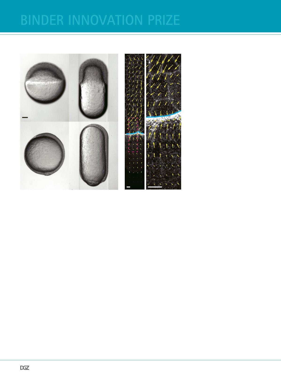
Figure 2:
Cable-constriction of the actomyosin band is not necessary for EVL epiboly.
a (a-b) Aspiration of Tg(actb2:myl12.1-eGFP) embryos into agarose tube deform embryos towards a
cylindrical shape. Brightfeld images were taken of control embryos and cylindrical embryos at 30%
epiboly (a,a’) and at 100% epiboly (b,b’). c) Actomyosin band formation, cortical fows and epiboly
progression are largely unaffected in cylindrical embryos at 60-70% epiboly. Cortical fow velocities are
quantifed using PIV (yellow arrows). Scale bars, 25 µm. Adapted from (15).
binder innovation prize


