cell news 2/2013
7
EVL
YSL
A
V
t=-1.2s
t=0s
t=3s
t=-1.2s
t=0s
t=3s
A
V
EVL
YSL
EVL
YSL
Myl12.1-eGFP
Lifeact-RFP
A
V
retrograde
flow
A
v
a
b
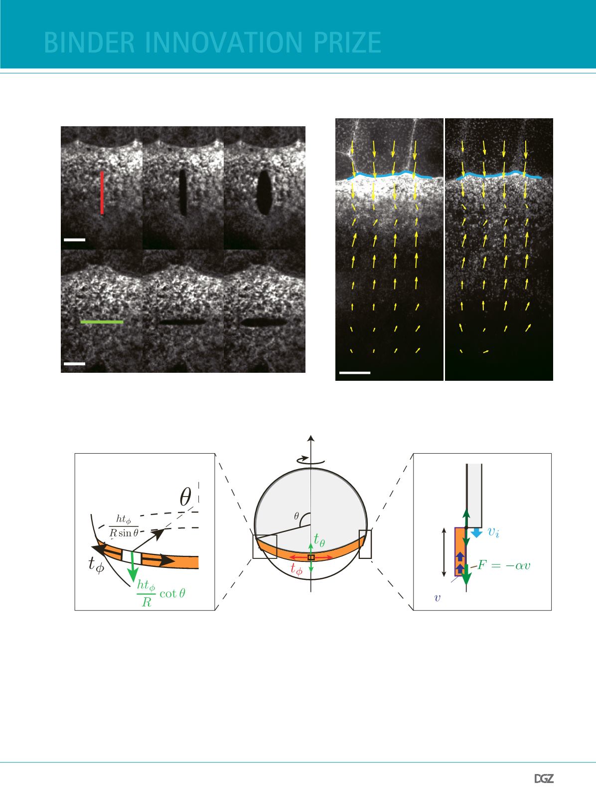
Figure 2:
Cortical tension and actomyosin fow within the YSL suggest two modes of actomyosin ring propulsion.
(a) Laser ablation experiments in the YSL actomyosin band of Tg(actb2:myl12.1-eGFP) embryos at 65% epiboly reveal substantial circumferential tension (red,
upper panel) as well as tension along the AV axis (green, lower panel). Scale bars, 10 µm.
(b) In Tg(actb2:myl12.1-eGFP) embryos injected with lifeact-RFP mRNA actomyosin band progression at 65% epiboly is accompanied by retrograde fow of
myosin (left) and actin (right) from vegetal parts of the yolk cell into the EVL margin (blue line). Particle image velocimetry (PIV) allows for quantifcation of
the fow velocity feld (yellow arrows). Scale bar, 20 µm.
(c) Theory of epiboly movements. Modeling the EVL and YSL tissues as thin active viscous layers reveal a two-fold contribution of the actomyosin ring to EVL
epiboly. (i) Active tension along the circumference of the actomyosin ring couples to the spherical geometry of the embryo resulting in a net force towards
the closest pole. (ii) Retrograde actomyosin fows are resisted by friction against a substrate and exert a pulling force onto the EVL tissue. Adapted from (15).
Vegetal Pole
Animal Pole
EVL
Actin
ring
Yolk
cell
R
-
Downward force
on tissue from
constriction
coupled to
geometry
(i) Cable constriction motor
(ii) Flow-friction motor
Retrograde
flow
Friction force
arising from
retrograde flow
pulls on tissue
h
c
binder innovation prize


