Cell News 2/2016
13
DGZ AWARD WINNERS 2016
where they are arrested in prophase of the first meiotic division.
Once every menstrual cycle, an oocyte resumes meiosis and com-
pletes the first meiotic division to mature into a fertilizable egg
(Fig. 1). First, the nucleus breaks down and a spindle assembles
in the center of the oocyte. In the next step, the spindle reloca-
tes from the oocyte’s center to its surface. When the spindle is
asymmetrically positioned, it segregates the homologous chromo-
somes and eliminates half of them in a small cell called polar body.
The remaining chromosomes become aligned in the metaphase II
spindle, and the egg stays arrested in this stage until it is fertilized.
Upon fertilization, the egg completes the second meiotic division,
in which it eliminates half of the non-identical sister chromatids
into the second polar body. The male and female pronuclei form
and the mitotic divisions of the embryo start.
We still know very little about the mechanisms that govern accu-
rate meiosis in mammalian oocytes, and it is still unclear what is
causing the high frequency of chromosome segregation errors in
human oocytes. The main aim of our laboratory is to investigate
how the oocyte’s cytoskeleton drives the many steps that are in-
volved in generating a viable and healthy embryo, and how defects
at the interface between chromosomes and cytoskeletal structures
lead to aneuploid eggs and pregnancy loss in mammals. To have
a solid foundation for future research in our laboratory, we are
developing new tools to study meiosis in mammalian oocytes. For
instance, we have been able to establish methods that now allow
us for the first time to study the causes of chromosome segregati-
on errors directly in live human oocytes.
Analysis of cytoskeletal organization and function in
oocyte meiosis
Many essential steps of meiosis are driven by cytoskeletal structu-
res. These cytoskeletal structures often differ from their seemingly
more reliable mitotic counterparts. Thus, differences in cytoskeletal
organization might contribute to meiotic errors. For instance, chro-
mosome segregation is driven by a specialized meiotic microtubule
spindle that – in contrast to mitotic spindles - lacks centrosomes
at its poles (Manandhar et al., 2005); and asymmetric positioning
of the spindle in mouse oocytes is actin- instead of microtubu-
le-dependent as in most mitotic cells (Longo and Chen, 1985). A
growing body of evidence suggests that actin and myosins also
have important functions for chromosome segregation and spind-
le assembly during meiosis in various organisms (Sandquist et al.,
2011). For instance, actin is required for chromosome congression
in starfish oocytes (Lenart et al., 2005) and a myosin is essential
for spindle assembly in Xenopus oocytes (Sandquist et al., 2011).
Whether actin and myosins have similar functions in mammalian
oocytes remains to be investigated.
Over the past few years, we have made significant progress in ana-
lyzing essential functions of actin in mouse oocytes. In addition, we
have studied how the microtubule spindle is organized in mamma-
lian oocytes and at the oocyte-to-embryo transition. This includes
the first studies of spindle assembly in live human oocytes.
Unexpected functions of actin in mammalian oocytes
Much of our work has focused on understanding how actin helps
to position the spindle asymmetrically before the extrusion of the
first polar body (Fig. 1). This is an important question because
asymmetric spindle positioning is a prerequisite for the extremely
asymmetric division of the oocyte, which ensures that the egg re-
tains sufficient storage material for embryo development.
Our previous work established that asymmetric spindle positio-
ning in mouse oocytes requires a cytoplasmic actin network that
is assembled by the actin nucleation factor Formin-2. The spindle
pulls on this network while it moves to the oocyte’s surface in a
myosin-dependent manner (Schuh and Ellenberg, 2008). We found
that Spire-type actin nucleation factors cooperate with Formin-2
to drive the asymmetric division of mouse oocytes (Fig. 2). In par-
ticular, Spire proteins and Formin-2 rely on each other to assemble
the cytoplasmic actin network that mediates asymmetric spindle
positioning and are essential for ingression of the cytokinetic fur-
row. Depletion of Spire proteins results in diploid eggs that cannot
give rise to euploid embryos upon fertilization (Pfender et al., 2011).
We further investigated the cooperation between Spire-type ac-
tin nucleation factors and Formin-2 in vitro in collaboration with
Marie-France Carlier’s group. This work revealed that Spire and
Formin-2 assemble F-actin by a ping-pong mechanism, in which
the actin filament is repetitively passed between the two proteins
during assembly (Montaville et al., 2014).
We have also investigated other functions of the actin network in
the oocyte. Surprisingly,
we found that it is requi-
red to transport vesicles
over long distances. In
particular, we found that
Rab11a-positive vesicles
move directionally along
the network in a myosin-
Vb-dependent manner to
converge and to reach
the cell surface. Micro-
tubules were dispensable
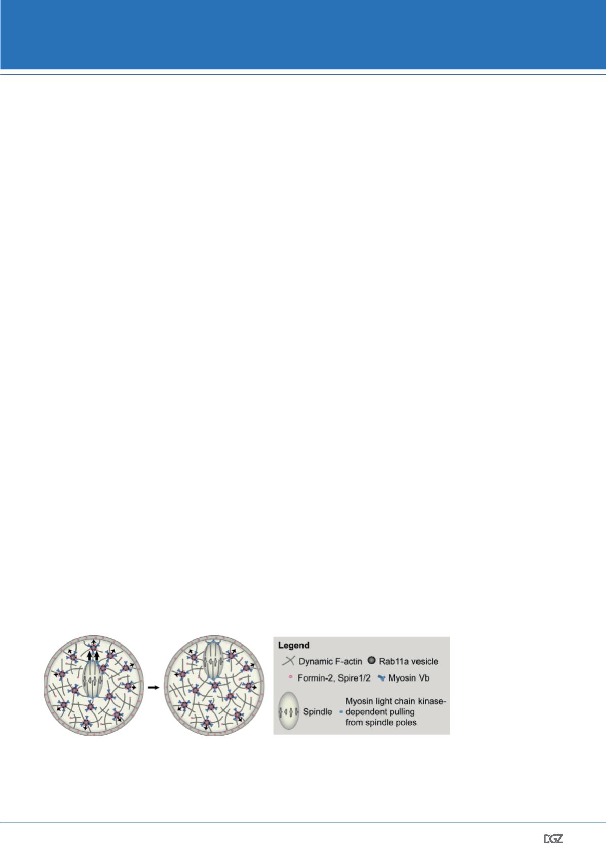
Figure 2. Mechanism of actin-dependent spindle and vesicle transport.
Past work in our group revealed that asymmetric spindle positioning is driven by a cytoplasmic actin network that is
nucleated by cooperation between the actin nucleation factors Formin-2 and Spire1/2. Spindle movement along this
network is facilitated by myosin-dependent pulling from the spindle poles and a outward-directed myosin Vb-dependent
movement of Rab11a-positive vesicles, which keep the network dynamic and help to modulate its density by localizing
and transporting the actin nucleation factors.
© Holubcova et al., Nat Cell Biol 2013


