Cell News 04/2019
27
LN capsule and subcapsular sinus. Perinodal lymphatics also
promote local interstitial flow, which cooperates with lympho-
toxin-
β
signaling to amplify stromal CXCL13 production and
thereby promote LTi cell retention and LN organogenesis.
Acknowledgements
Although only my name appears in this review, I would like to
acknowledge all the co-authors who made this story possible. I
am grateful for their work, intellectual input and great discus-
sions. A special thank to Prof. Tatiana Petrova, whose passion
for science and strong support were a great motivation during
the last years. A special thank to Dr. Amélie Sabine, for her
enthusiastic supervision and tremendous support. I also would
like to thank all members of the Petrova group for their help
and amazing working environment. Finally, I am thankful to the
Werner Risau Prize committee and the DGZ for recognizing this
work. It is a privilege to be part of the vascular biology commu-
nity, pioneered by Werner Risau.
About the author
Esther Bovay (PhD)
Max Planck Institute for Molecular
Biomedicine,
Department of Tissue Morphogenesis,
Röntgenstraße 20, 48149 Münster,
Germany
E-mail:
03.2019 to present: Postdoctoral researcher
Prof. Ralf Adams laboratory, Max Planck Institute for Molecular
Biomedicine, Münster, Germany
03.2013 – 02.2018: PhD student
Prof. Tatiana Petrova laboratory, Department of Oncology, Cen-
tre Hospitalier Universitaire Vaudois and University of Lausanne,
Epalinges, Switzerland
09.2011 - 01.2013: M.Sc Biology
University of Lausanne, Switzerland
09.2008 - 06.2011: B.Sc Biology
University of Lausanne, Switzerland
References
Ansel, K.M., Ngo, V.N., Hyman, P.L., Luther, S.A., Förster, R., Sedg-
wick, J.D., Browning, J.L., Lipp, M., Cyster, J.G., 2000. A chemo-
kine-driven positive feedback loop organizes lymphoid follicles.
Nature 406, 309–314.
Brendolan, A., Caamano, J., 2012. Mesenchymal cell differen-
tiation during lymph node organogenesis. Front. Immunol. 3.
Bromley, S.K., Thomas, S.Y., Luster, A.D., 2005. Chemokine re-
ceptor CCR7 guides T cell exit from peripheral tissues and entry
into afferent lymphatics. Nat. Immunol. 6, 895–901.
org/10.1038/ni1240
Carrasco, Y.R., Batista, F.D., 2007. B cells acquire particulate
antigen in a macrophage-rich area at the boundary between the
follicle and the subcapsular sinus of the lymph node. Immunity
27, 160–171.
Chambliss, A.B., Khatau, S.B., Erdenberger, N., Robinson, D.K.,
Hodzic, D., Longmore, G.D., Wirtz, D., 2013. The LINC-anchored
actin cap connects the extracellular milieu to the nucleus for
ultrafast mechanotransduction. Sci. Rep. 3, 1087.
org/10.1038/srep01087
Cohen, J.N., Guidi, C.J., Tewalt, E.F., Qiao, H., Rouhani, S.J., Rud-
dell, A., Farr, A.G., Tung, K.S., Engelhard, V.H., 2010. Lymph node–
resident lymphatic endothelial cells mediate peripheral tolerance
via Aire-independent direct antigen presentation. J. Exp. Med.
207, 681–688.
Cordeiro, O.G., Chypre, M., Brouard, N., Rauber, S., Alloush, F.,
PRIZE WINNERS 2019
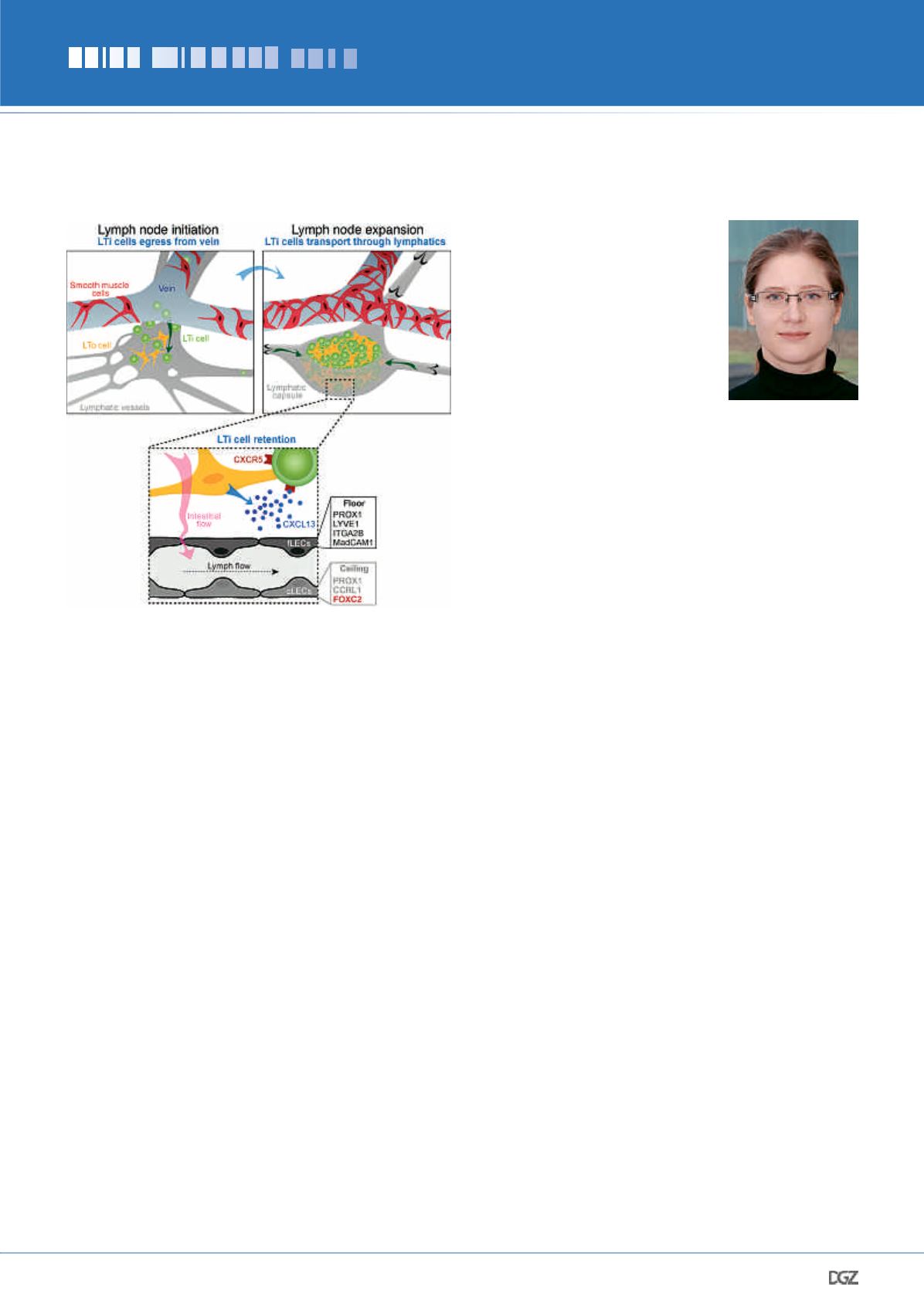
Figure 8. Lymphatic vessels in LN development.
LTi cells extravasate from the immature vein to initiate LN formation.
At the same time, lymphatic vessels take up disseminated LTi cells, ex-
travasating from blood capillaries elsewhere, and transport them to the
LN anlage. Later, collecting vessels efficiently transport LTi cells to LNs
and participate in their expansion. Collecting vessels reorganize into a
LN capsule, which engulfs the LN anlage. LN lymphatic fluid absorption
from blood vessels generates IFF, which increases CXCL13 expression,
improving LTi cell retention.


