Cell News 04/2019
17
PRIZE WINNERS 2019
between epithelial (hepatocytes and ductal cells) endothelial
(sinusoidal cells) and mesenchymal cells (portal fibroblasts and
stellate cells) (11).
During organogenesis, liver embryonic progenitor cells (known as
hepatoblasts) are specified from the posterior foregut endoderm.
In response to signalling factors secreted by the surrounding
mesenchyme, such as FGF, BMP, HGF and Wnt, hepatoblasts
undergo cell shape changes, proliferate and migrate into the ad-
jacent mesoderm to form the liver bud(12). During the course of
liver bud outgrowth, hepatoblasts become lineage committed in
order to give rise to hepatocytes and cholangiocytes(13). Indeed,
we recently reported that a single
Lgr5+
hepatoblast can gener-
ate both hepatocytes and cholangiocytes, demonstrating for the
first time that single hepatoblasts are bipotent (10). The fate of
hepatoblasts is influenced by local signalling: subsets of hepa-
toblasts that are exposed to signals near the portal mesenchyme
generate cholangiocytes, whilst hepatoblasts that are located
further from the portal veins respond to signals from closely
associated haematopoietic cells and give rise to hepatocytes.
To support normal functions, the adult liver must be maintained
during homeostasis. In contrast to other endodermal organs such
as the intestine that self-renew every 3-5 days, the liver has a
much slower cellular turnover, (in mice, approximately every 60
and 150 days for cholangiocytes and hepatocytes, respective-
ly (14)). Homeostatic epithelial maintenance occurs primarily
through the self-duplication of mature cells(15-16). Despite a
low cellular turnover, when challenged, the liver has a remark-
able ability to regenerate, although repeated damage to the
tissue can result in impairment of liver function and fibrosis, as
reviewed in (17). Upon partial hepatectomy (surgical resection of
up to 2/3 of the liver) the remaining healthy mature hepatocytes
respond to injury-induced regenerative signals such as TNFa and
interleukin (IL)-6 to proliferate and undergo hyperplasia in order
to restore tissue mass within a week(18-19). Understanding of
this phenomenon has been taken into the clinic and helped to
facilitate live-donor transplants and tumour resections. How
ever, upon toxin-mediated damage, (e.g. viruses and alcohol) or
due to chronic liver pathologies such as non-alcoholic fatty liver
disease (NAFLD), hepatocytes become impaired and are unable
to undergo the mass proliferative response seen following partial
hepatectomy. Incredibly, even when hepatocyte proliferation is
compromised the liver is still capable of regenerating itself. In
this case, there is a ductular reaction in which duct cells become
activated and start to proliferate, repopulating the liver (10-
23). Understandably, there has been a large effort to establish
faithful
in vitro
liver models to gain insights not only into liver
biology and diseases but also into regenerative mechanisms in
general (Figure 2). Below I summarize our contribution to this
knowledge.
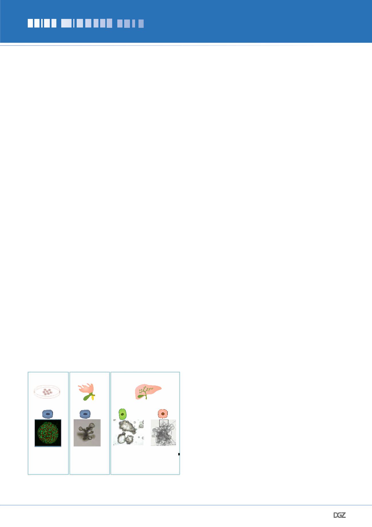
Organoids that recapitulate liver tissue and liver
regeneration
For a long time, in vitro expansion of adult hepatocytes and/
or cholangiocytes remained a challenge. Pioneering work from
Michalopoulos et al. had shown that primary liver cells cultured
in 3D could be maintained in culture in the presence of EGF,
HGF and Dexamethasone (24). However, these conditions did not
allow the long-term expansion of liver cells ex vivo. We utilized
Matrigel as a 3D basal ECM collagen and laminin-rich matrix in
combination with a cocktail of growth factors known to play a
role in liver development and/or regeneration to establish the
first liver organoid model as we know it today.
To establish liver organoid cultures, our first approach was to
gain further understanding on how the adult mouse liver cells
activate a proliferative program during regeneration so we could
recapitulate in vitro this pro-regenerative growth-factor envi-
ronment. Our mouse in vivo studies indicated that adult liver ac-
tivates Wnt signaling upon liver damage (8), hence our approach
started by boosting Wnt signaling by addition of R-spondin1, a
Wnt agonist essential for mouse small intestinal (4) and stom-
ach (5) cultures and later found to be the ligand for
Lgr5
(25).
Next, we added the mitogens EGF and FGF10, as FGF7/FGFR2-
signalling, required for the expansion of mouse liver ductal cells
in vivo, during regeneration (26). Hence, we defined a mouse
liver organoid medium containing the cocktail of growth factors
Egf, Rspo1, FGF10, HGF and Nicotinamide, which would support
the long-term expansion of mouse liver cells even from a single
isolated ductal or
Lgr5
+
cell. By combining this cocktail with
embedding the cells in lamini-rich ECM, we found that isolated
mouse liver cells (healthy liver ducts or
Lgr5
+
liver cells post
damage-induction) self-organized into 3D structures that retain
the ability to differentiate into functional hepatocyte-like cells
in vitro
(8) and also
in vivo
upon transplantation into a mouse
model of tyrosinemia type I liver disease. Of note, we recently
described the isolation of bipotent
Lgr5
+
embryonic hepatoblasts
which retain the capacity to form either hepatocyte or cholan-
Figure 2: Liver organoid cultures. Chol, cholangiocyte (also known as
duct cell). Hep, hepatocyte.
iPSCs
Takebe et al. Nature 2013
Takebe Cell Rep 2017
Sampaziotis Nat Biotech
2015
Embryonic Liver
Hepatoblast
Hu et al. Cell 2018
Prior et al Development
2019
Hepatocyte-like
cell
Adult Liver
Hepatocytes
Duct cells
Huch et al. Nature 2013
Huch et al. Cell 2015
Broutier et al. Nat Prot 2016
Hu et al. Cell 2018
Peng et al., Cell 201
8
Liver Bud and Chol
organoids
Hep and Chol
Embyonic organoids
Chol-derived
organoids
Hep-derived
organoids


