Cell News 04/2019
9
PRIZE WINNERS 2019
inner and an outer SVZ (iSVZ and oSVZ, respectively) (Cárdenas
and Borrell, 2019; de Juan Romero et al., 2017; Fernandez et al.,
2016), consequently augmenting in general the size and total
surface area of the brain.
Correcting an almost 20 years old mistake: Akna
is a novel cell type specific centrosomal protein
We searched for candidate regulators of murine cortical NSCs
involved in SVZ generation and expansion (Pinto et al., 2008;
Pinto et al., 2009; Stahl et al., 2013), and found a promising
gene called Akna. We chose to examine the role of this putative
AT-hook containing transcription factor (Siddiqa et al., 2001) as
we found it to have elevated mRNA expression in differentiating
NSCs (generating BPs) while self-renewing NSCs have lower
expression levels, and because its mRNA correlated with the
time at which the SVZ is generated in mice (i.e. low at E11, high
at E14 and low at E18). We were very surprised to observe cen-
trosomal localization of Akna (Figure 2) in a variety of cell types
(including immune cells, where it was initially characterized) of
murine, ferret and primate origin, but no nuclear localization.
This was done by means of immunofluorescent staining using
several monoclonal antibodies generated by us, and subsequent-
ly validated through other methods including superresolution
and electron microscopy, mass-spectrometry of purified centro-
somes, protein tagging, BAC-transgenomics (Poser et al., 2008)
and RNA-interference. Our observation is also in agreement with
Jakobsen et al., 2011, where they detected Akna at centro-
somes but did not investigate its role. Akna is an
integral part
of subdistal appendages (SDAs) of centrosomes as its centroso-
mal localization is not altered by disruption of microtubules or
Dynein-motor complexes, but only by elimination of centrioles
or SDAs.
We found out that the cDNA clone used in the original publi-
cation lacked almost half of the N-terminal domain (containing
the MT-binding domain), while a large part of the C-terminal
region (containing the centrosomal targeting domain) did not
correspond to Akna. The cDNA clone used by Siddiqa and col-
leagues was therefore compromised and very likely not function-
al. Furthermore, we found that the AT-hook (the supposed DNA
binding domain) was not conserved among vertebrates, not even
within near related primates (see supplementary discussion in
Camargo Ortega et al., 2019).
Microtubules dynamics regulate NSCs delamination
We asked next what is the function of Akna in NSCs and BPs.
Towards this end we first checked the distribution of Akna
protein in the developing cortex. In complete agreement to the
mRNA profile, Akna was present at high levels at the centro-
somes of differentiating NSCs and BPs in the SVZ, and absent
in proliferating (non-differentiating) NSCs and neurons in the
CP (Figure 3a). To our knowledge, Akna is the only centrosomal
protein described so far with such cell-type specific expression
in the developing brain. Next, we performed gain- and loss-
of-function experiments in NSCs by electroporating plasmids
encoding Akna cDNA or anti-Akna shRNAs, respectively.
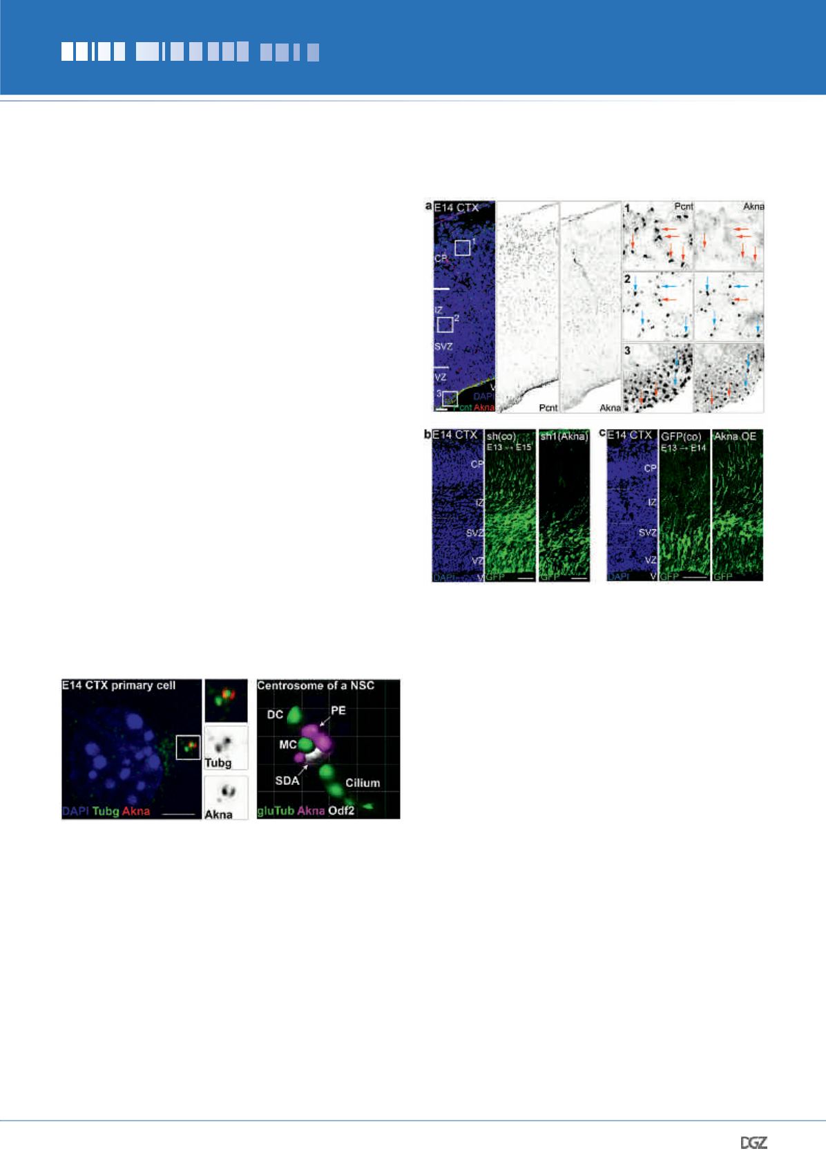
Figure 2:
Akna is a centrosomal protein. The figure on the left shows
the co-localization of Akna with the centrosomal protein Tubg (gamma
Tubulin) in a cell the murine cerebral cortex at E14. Notice the enrich-
ment around one of the centrioles. The figure on the right is a magni-
fication of the centrosome of an E14 CTX NSC showing the localization
of Akna at SDAs (co-localization with Odf2) and at PEs. CTX, cortex;
NSC, neural stem cell; PE, proximal end; SDA, sub-distal appendage.
Bar = 5 μm.
Figure 3:
Expression and function of Akna in NSCs of the developing
CTX. (a) Akna is a centrosomal protein specifically expressed in differ-
entiating NSCs in the VZ and BPs in the SVZ, and absent in proliferating
NSCs and neurons in the CP (red arrows). (b) Akna knockdown with
shRNAs impairs the delamination and migration of NSCs to the SVZ.
(c) Akna overexpression in NSCs promotes a fast delamination of NSCs
towards the SVZ. Bars = 20 μm (a), 50 μm (b,c).


