Cell News 04/2019
8
PRIZE WINNERS 2019
The intricate, yet exquisitely organized shape, as well as the
particular large size, are the most notorious features of our
brain. This has contributed to the acquisition of evolutionary ad-
vantages und ultimately to the superposition of humans at the
top of cognitive beings (Cárdenas and Borrell, 2019; Fernandez
at al., 2016; Klyachko and Stevens, 2003). Understanding how
our brain forms and acquires its final architecture is therefore
fundamental, in one hand, to learn about the etiology of devel-
opmental diseases and, eventually, how to prevent or ameliorate
them. In the other hand, for the efficient manipulation of the
brain’s cellular components and future use of these in regener-
ative medicine. Our laboratory spearheads the research on novel
cell biological mechanisms regulating the self-renewal and dif-
ferentiation of the progenitor cells in the developing and adult
mammalian brain (Beckervordersandforth et al., 2010; Camargo
Ortega et al., 2019; Ninkovic et al., 2013; Pinto et al., 2008;
Pinto et al., 2009; Stahl et al., 2013). This has led to the discov-
ery and pioneering development of protocols to direct neuronal
reprogramming of nonneuronal cells and repair in the injured
brain (Gascón et al., 2016; Masserdotti et al., 2015; Ninkovic
et al., 2013; Mattugini et al., 2019). Here, I will describe the
discovery and characterization of a novel and unexpected factor
controlling early differentiation steps of embryonic neural pro-
genitors and discuss the broader relevance of our studies.
Neurogenesis in the developing mammalian
cerebral cortex
Neural stem cells (NSCs) of the developing mammalian cerebral
neocortex are neuroepithelial radial glia cells (Götz, el al., 2016;
Götz and Huttner, 2005; Kriegstein and Alvarez-Buylla, 2009)
and generate most, if not all, pyramidal excitatory neurons, as
well as nonneuronal cells such as astrocytes, ependymas cells,
oligodendrocytes (Götz et al., 2016; Kriegstein and Alvarez-Buyl-
la, 2009) and the future NSCs of the adult brain (Falk and Götz,
2017) (Figure 1). In the mouse, embryonic cortical neurogenesis
peaks at around embryonic day 14 (E14). At this point, NSCs that
have committed to differentiation delaminate from their niche
at the ventricular zone and move basally where they transform
into intermediate transient amplifying basal progenitors (BPs).
They do so by retracting both apical and basal processes and
acquiring a characteristic
multipolar
morphology. They also
re-position cytoskeletal components such as centrosomes and
microtubule networks, and this is required to re-orient the cell
soma as they further differentiate into
bipolar
neurons that later
migrate out of the SVZ, pass through the intermediate zone
(IZ), and seed the cortical plate (CP) (Figure 1). BPs undergo
an additional round of cell division with the goal of increasing
the number of future neurons, therefore and thereby forming
a secondary germinal subventricular zone (SVZ). The time that
BPs reside in the SVZ is important to permit major cellular and
genetic changes to happen. Importantly, in species with a folded
brain, such as ours, the SVZ is expanded and subdivided into an
Nikon Young Scientist Award
Centrosome and microtubule dynamics regulate the balance
of stem cell self-renewal and differentiation
University of Munich, Department of Physiological Genomics; Helmholtz Zentrum München,
Institute of Stem Cell Research; ETH Zürich, Department of Biosystems Science and Engineering
Germán Camargo Ortega
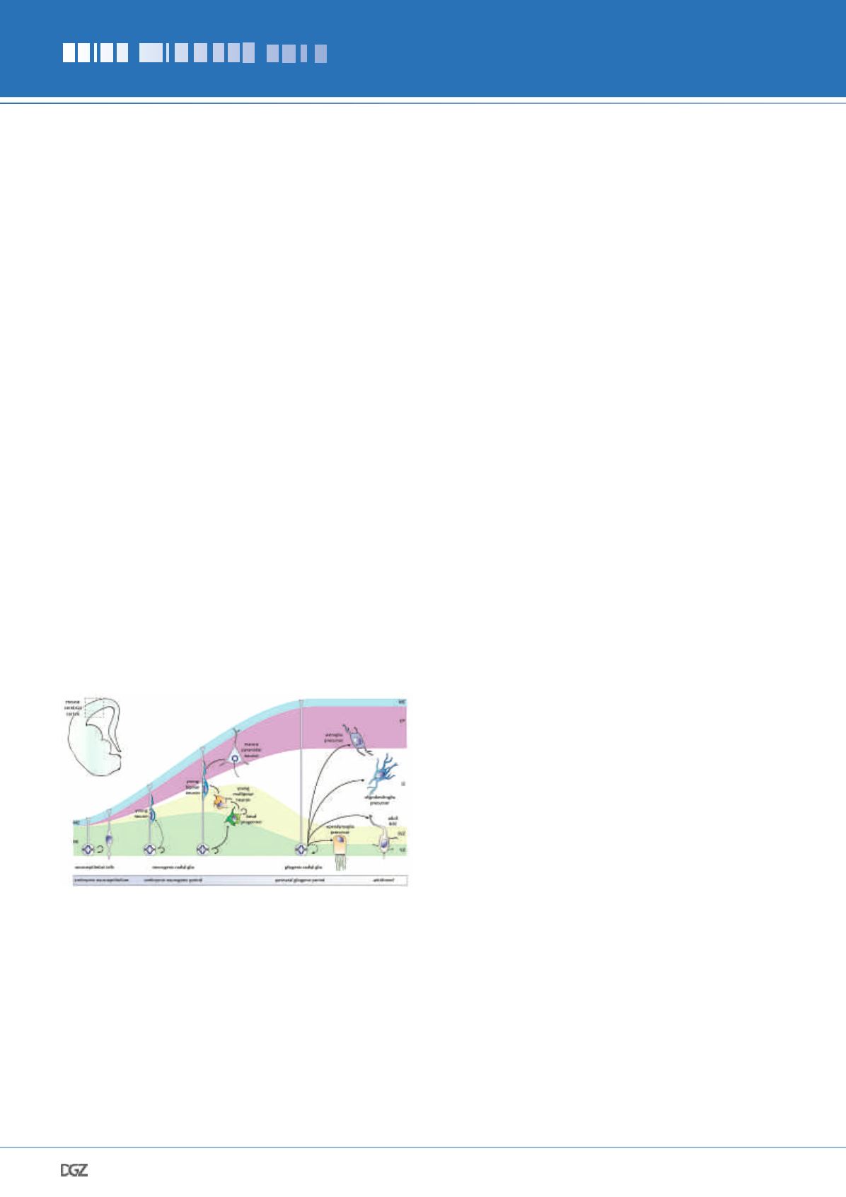
Figure 1:
Development of the cerebral cortex. NECs at the VZ divide
symmetrically and amplify the progenitor pool and subsequently trans-
form into NSCs (a.k.a. radial glia) that both self-renew and generate
neurons directly or indirectly via intermediate BPs. The latter increase
the neuronal output after additional rounds of cell division, thus
forming a secondary germinal layer; the SVZ. Neurogenesis reaches
its end nearing perinatal stages, while glia cells start to be generated:
ependymo-, astro- and oligodendrogliogenesis occurs sequentially.
A subpopulation of RGCs is separated during neurogenesis, becomes
quiescent and transforms later into adult NSCs. BP, basal progenitor;
CP, cortical plate; IZ, intermediate zone; MZ, marginal zone; NE, neu-
roepithelium; NEC, neuroepithelial cell; SVZ, subventricular zone; VZ,
ventricular zone.


