20
Cell News 2/2015
cular level is an efficient way to discover genes contributing to
specific barrier functions. With this approach we discovered a no-
vel gene important in creating an impermeant BBB by controlling
vesicular transport across the barrier, an attractive path for drug
delivery.
In order to identify mechanisms governing the establishment of
a functional BBB, we first developed a novel tracer-injection me-
thod for embryos and demonstrated spatiotemporal developmen-
tal profiles of BBB functionality. The prevailing view has been that
the embryonic and perinatal BBB are not yet functional
1
. Howe-
ver, previous embryonic BBB functionality studies were primarily
performed by trans-cardiac tracer perfusion, which may dramati-
cally affect blood pressure, cause bursting of CNS capillaries, and
artificially produce leakiness phenotypes
1
. To circumvent these
obstacles, we developed a method to assess BBB integrity during
mouse development, in which a small volume of tracer is injected
into embryonic liver to minimize changes in blood pressure (Fig.
1a). With this method we found that the mouse BBB becomes
functional at embryonic day 15.5 (E15.5 Fig. 1b). Specifically, at
E13.5 cortex a 10-kDa dextran tracer leaked out of capillaries and
was taken up by non-vascular brain parenchyma cells (Fig. 1b, top
panel). At E14.5, the tracer was primarily restricted to capillaries,
but tracer was still detected outside vessels (Fig. 1b, middle pa-
nel). In contrast, at E15.5, the tracer was confined to vessels with
no detectable signal in the surrounding brain parenchyma, similar
to the mature BBB (Fig. 1b, bottom panel). These data demonstra-
te that following vessel ingression into the neural tube, the BBB
gradually becomes functional as early as E15.5.
Identifying the developmental time-point when the BBB gains
functional integrity, enabled us to use that time window to profi-
le BBB-specific genes when the BBB is actively forming. Based on
the temporal profile of BBB formation, we compared expression
profiles of BBB (cortex) and non-BBB (lung) endothelium at E13.5,
using an Affymetrix array, and identified transcripts with signifi-
cantly higher representation in cortical than lung endothelium.
These transcripts included transporters, transcription factors, and
secreted and transmembrane proteins. We were particularly in-
terested in transmembrane proteins, owing to their potential in-
volvement in cell–cell interactions that regulate BBB formation.
One of the genes identified,
Mfsd2a
, had 78.8 times higher ex-
pression in cortical endothelium than in lung endothelium (fig.
2a). Both
in situ
hybridization and Immunohistochemistry showed
prominent expression in CNS vasculature but no detectable signal
in vasculature outside the CNS, such as in lung or liver. Moreo-
ver, both
Mfsd2a
mRNA and
MFSD2A
protein were absent in the
choroid plexus vasculature, which is part of the CNS but does not
possess a BBB
1
(Fig. 2c, d, g).
MFSD2A
expression in CNS vascula-
ture was observed at embryonic stages (E15.5), postnatal stages
(P2 and P5) and in adults (P90). Finally,
MFSD2A
protein, which is
absent in the
Mfsd2a
-\-
mice (Fig. 2e)
15
, was specifically expressed
in claudin-5-positive CNS endothelial cells but not in neighbou-
ring parenchyma cells (neurons or glia) or adjacent pericytes (Fig.
2f). Previously,
MFSD2A
was reported to be a transmembrane
protein expressed in the placenta and testis, which have highly
restrictive barrier properties
16,17
.
Genetic ablation of
Mfsd2a
resulted in a leaky BBB from emb-
ryonic stages through to adulthood, suggesting that
Mfsd2a
is
critical for the formation and function of the BBB. While barri-
er genesis is greatly affected in these mice, this
Mfsd2a
genetic
ablation resulted in the normal patterning of vascular networks,
suggesting that
Mfsd2a
is critical for barrier-genesis but not for
the CNS angiogenic program. Using our embryonic injection me-
thod, 10-kDa dextran was injected into
Mfsd2a
-\-
and wild-type
littermates at E15.5. As expected, dextran was confined within
vessels of control embryos. In contrast, dextran leaked outside the
invaginated from the luminal membrane and exocytosed at the ablum-
inal plasma membrane only in
Mfsd2a
2
/
2
mice (Fig. 5d), suggesting
that HRP was subject to transcytosis in these animals but not in wild-
type littermates (ExtendedDataTable 2). Together, these findings suggest
that the
opening
ficking a
Studie
that peri
vesicle tr
servation
that Mfs
pericyte f
tosis is m
capillary
endotheli
These da
suggest t
lial cells i
reductio
d
b
*
5,000
4,000
3,000
2,000
1,000
a
500 µm
c
g
P5
Mfsd2a
0
e
P5
P2
f
Pdgfr
β
**
*
E15.5
E15.5
P5
Lectin
Mfsd2a
Mfsd2a/Claudin-5/DAPI
Cortex
Lung
PECAM
PECAM
Mfsd2a
Mfsd2a/Claudin-5
Claudin-5
Mfsd2a/
Claudin-5
Mfsd2a
Mfsd2a/
Pdgfr
β
Expression (a.u.)
500 µm
E15.5
Mfsd2a
100 µm
100 µm
100 µm
5 µm
100 µm
Mfsd2a +/+
Mfsd2a
–/–
Mfsd2a
+/+
Mfsd2a +/+
a
10-kDa tracer
Lectin
Overlay
b
E15.5
Percentage of sample
0
100
80
60
40
20
0
0
0.5
1
1.5
2
2.5
Spectrophotometric values
(fold change)
WT MUT
P90
*
c
Mfsd2a –/–
Mfsd2a –/–
25 μm
25 μ
Tracer-filled parenchyma cells
5-10
1-5
Figure 4
|
Mfsd2a
is required for the establishment of a functional BBB but
not for CNS vascular patterning
in vivo
. a
,
b
, Dextran-tracer (10 kDa)
injections at E15.5 revealed a defective BBB in mice lacking
Mf d2a
.
a
, The
tracer was confined to the capillaries (arrow) in wild-type littermates, whereas
Mfsd2a
2
/
2
embryos showed large amounts of tracer leakage in the brain
parenchyma (arrowheads).
b
, Capillaries (arrows) surrounded by tracer-filled
brain parenchyma cells (arrowheads) in
Mfsd2a
2
/
2
cortex. Quantification of
tracer-filled parenchy a cells in control versus
Mfsd2a
2
/
2
cortical plates
(bottom panel,
n
5
7 embryos per genotype).
c
, Spectrophotometric
quantifica
post intra
persists in
exhibit no
vascular d
Quantific
genotype)
wild type.
Figure 3
|
vasculatu
,
80-fold
b–d
, Spec
Mfsd2a in
sections).
brain and
c
,
Mfsd2a
example,
plexus, da
in BBB-co
plexus (lef
e–g
, Imm
expressio
(endotheli
expressio
Mfsd2a
2
/
positive e
but not in
double ast
(fourth ve
Mfsd2a ex
Macmillan Publishers Limited. All ri
©2014
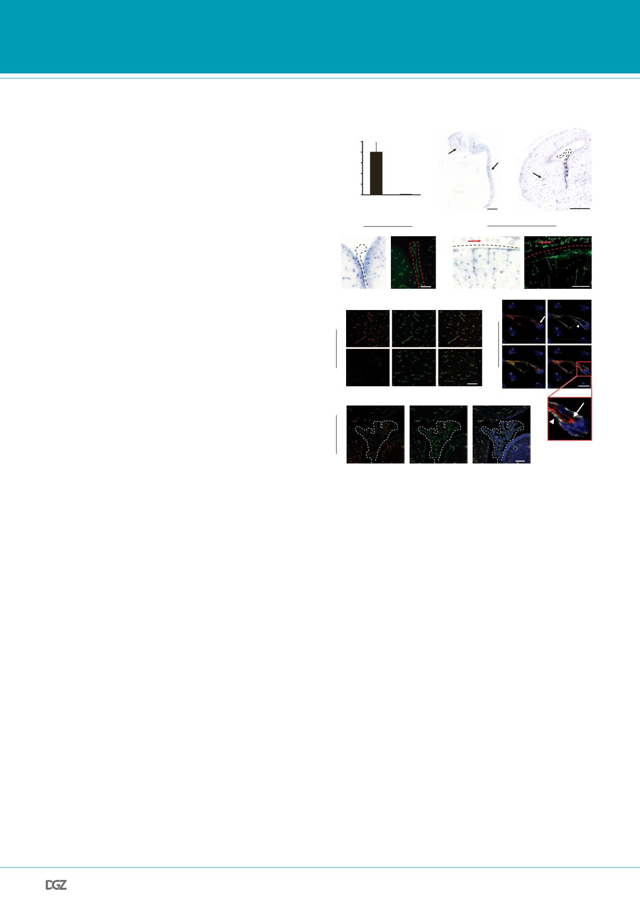
Figure 2 |
Mfsd2a
is selectively expressed in BBB-containing CNS
vasculature:
a, E13.5
Mfsd2a
expression in cortical endothelium was
~80-fold higher than lung endothelium (microarray analysis, mean±s.d.).
b-d, Specific
Mfsd2a
expression in BBB-containing CNS vasculature (Blue:
Mfsd2a
in situ
hybridization, green: vessel staining (PECAM) adjace t
sections). b, xpression in CNS vasculature (E15.5 sagittal view-brain and
spinal cord, arrows), but not in non-CNS vasculature (asterisk). c,
Mfsd2a
expression in BBB vasculature (E15.5 cortex coronal view e.g. striatum, ar-
row), but not in non-BBB CNS vasculature (choroid plexus, dotted line). d,
High magnification coronal view of
Mfsd2a
expression in BBB-containing
CNS vasculature but not in vasculature of the choroid plexus (left, dotted
line), or outer meninges/skin (right, red arrows). e-g, Immunohistochemi-
cal staining of
MFSD2A
protein shows specific expression in CNS endo-
thelial cells (Red:
MFSD2A
; green: Claudin5 or Lectin (endothelium); blue:
DAPI (nuclei); gray: PDGFR
β
(pericytes)). e,
MFSD2A
expression in the brain
vasculature of wild-type mice (upper panel), but not of
Mfsd2a
-/- mice
(lower panel). f,
MFSD2A
expression only in Claudin5-positive endothelial
cells (arrow, endothelial nucleus–asterisk) but not in adjacent pericytes (ar-
row head, pericyte nucleus–double asterisk). g, Lack of
MFSD2A
expression
in choroid plexus vasculature (fourth ventricle coronal view-dotted line),
as opposed to the prominent
MFSD2A
expression in cerebellar vasculature.
n=3 embryos (3 litters/age).
PRIZE WINNERS


