cell news 2/2013
25
research news
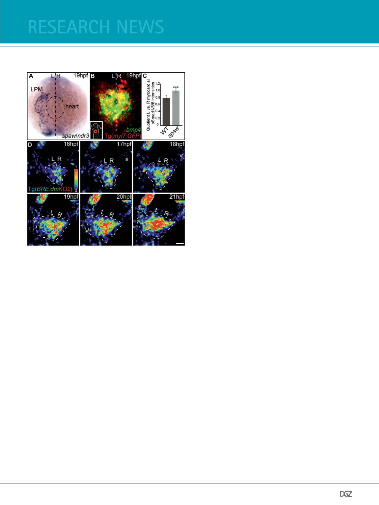
verse asymmetry of activity (Fig. 2C,D). Transgenic Bmp response
element (BRE) reporter fsh (Collery and Link, 2011) indicated
that the intensity of pSmad-1/5/8 activity (those pSmads are
activated by the Bmp signaling cascade) is signifcantly higher
on the right side of the cardiac cone (Veerkamp et al., 2013;
Fig. 2D). Hence, both Nodals and Bmps establish complementary
asymmetries of activity within the cardiac feld.
Such a complementary pattern could be due to an antagonism
between the two signaling cascades. Three lines of evidence
support the hypothesis that left-sided Spaw has an inhibitory
effect on Bmp activity: First, the loss of Spaw abolishes L/R dif-
ferences in Bmp signaling activity. Second, the misexpression of
Spaw in single myocardial cells suppresses Bmp signaling acti-
vity. Finally, in converse experiments, the loss of Bmp activity
in myocardial single cells does not infuence the expression of
Nodal target genes within the heart feld. These and other func-
tional tests suggested that Nodal signaling negatively affects
the activity of Bmp within the cardiac feld, raising the question
of the mechanism by which it does so.
In principle, any mechanism by which Spaw biases Bmp signa-
ling activity should also involve Spaw targets that affect cardiac
laterality. Candidate gene approaches revealed that the negative
modulation of Bmp signaling activity by Spaw is mediated by
the Nodal target Hyaluronan synthase 2 (Has2), an extracellular
matrix (ECM)-modifying enzyme which is asymmetrically ex-
pressed within the cardiac cone and which is required for car-
diac laterality (Smith et al., 2008; Veerkamp et al., 2013). The
enzyme Has2 is required for the production of hyaluronic acid,
an important ECM component that becomes cross-linked with
various proteoglycans. Clonal misexpression of Has2 within sin-
gle cells of the cardiac feld results in a signifcant local dam-
pening of Bmp signaling activity. Hence, Has2 has a local effect
on Bmp signaling activity, implying that the local hyaluronic
acid-proteoglycan composition of the ECM is inhibitory for au-
tocrine Bmp signaling. In part, such an effect could be due to
a scavenger function of these ECM components for bioactive
Bmp ligands on the left side of the cardiac feld.
bmp promotes epithelial and antimotogenic states
among cardiac progenitor cells
During zebrafsh cardiac development, the behavior of cardiac
progenitor cells appears to be tightly controlled by Bmp activity
and to shift from a non-motile epithelial state to motile mesen-
chymal-like states. Cell shape analyses combined with quanti-
fcations of cardiac progenitor cell motility rates revealed that
high levels of Bmp activity correlate with more epithelial, less
motile properties. Comparative microarray expression analyses
using highly enriched cardiac tissue helped to identify the Bmp
target genes involved in regulating these cellular properties.
Many genes that are positively regulated by Bmp encode cell
adhesion factors or determinants of epithelial character. A par-
ticularly intriguing target gene of Bmp is encoding nonmuscle
myosin II (NMII), important in epithelial remodeling, cellular
motility, and cell polarity (Conti and Adelstein, 2008; Widmann
and Dahmann, 2009; Lecuit et al., 2011). Consistent with po-
sitive regulation by Bmp, higher levels of phosphorylated NMII
(the activated form of this motor molecule) are present on the
right side of the cardiac cone. Both loss- and gain-of-func-
tion approaches for NMII activity revealed that NMII is indeed
an important regulator of cardiac laterality. Thus, cardiac L/R
asymmetry can partly be explained by an antimotogenic Bmp
activity that controls levels of NMII, thereby affecting both
cell shape and cell motility. In turn, Bmp activity is modula-
ted by asymmetrically expressed Nodal, which conditions the
ECM composition within the left cardiac feld (Veerkamp et al.,
2013; Fig. 3).
generating invariant organ form involves
random cell
motility gradients
Remarkably, when single cells misexpress Spaw, dominant-ne-
gative Bmp receptor, or constitutively-active Myosin light chain
Figure 2:
The L/R asymmetry of Bmp signaling activity depends on Nodal. (A) Whole-mount
in situ hybridization shows that expression of
spaw/ndr3
is restricted to the left
lateral plate mesoderm (LPM) neighboring the heart cone. (B) In comparison,
bmp4
(labeled green) is expressed throughout the entire heart cone (false-colored
in red) as shown by fuorescence two-color in situ hybridization. (C) The L/R
asymmetry of pSmad-1/5/8 intensities is abolished upon loss of Spaw/Ndr3. (D)
Despite symmetrical
bmp4
gene expression, Bmp signaling activity is higher on
the right side of the cardiac feld (outlined by white dotted line), as indicated by
the Bmp reporter line Tg(
BRE-AAVmlp:dmKO2
)
mw40
(Veerkamp et al., 2013)


