cell news 2/2013
19
mation of actin flaments. Compared to other so far identifed
actin nucleators such as Formins or Spire the the Arp2/3 com-
plex mainly initiates actin flaments on the sides of preexis-
ting mother flaments resulting in branched networks of actin
flaments enriched near the leading edge of cells (for review:
(Goode and Eck, 2007; Chesarone and Goode, 2009). This coup-
ling of actin nucleation and flament branching establishes the
basis of the dendritic nucleation model, which implies repeated
cycles of branching nucleation of actin flaments by the Arp2/3
complex generating the forces to push cell membranes (Mullins
et al., 1998). Improved cyro-electron tomographs confrmed the
existence of branched actin flaments, however these flaments
of variable length are not concentrated at the front but rather
distributed throughout protruding lamellipodia (Vinzenz et al.,
2012). Thus, the authors proposed that branching might be im-
portant for generating an actin network, but force generation
is not dependent on short flaments generated at branch points
(Vinzenz et al., 2012, Small et al., 2008). This also implies a new
model for membrane protrusion that requires an unknown cros-
stalk between different actin nucleating, elongating and cross-
linking complexes in vivo.
Wiskott-Aldrich syndrome protein (WASP) family
members – key regulators of the actin cytoskeleton
The activity of the Arp2/3 nucleation machine is controlled by
so-called nucleation promoting factors (NPF) such as mem-
bers of the Wiskott-Aldrich syndrome protein (WASP) fami-
ly that drive actin polymerization in time and space (Derivery
and Gautreau). In vertebrates, the WASP/WAVE protein family
consists of eight different proteins: the two Wiskott-Aldrich
syndrome proteins WASP and N-WASP, the related WASP fami-
ly Verprolin homologous proteins WAVE 1-3 (also called SCAR
1-3), the Wiskott-Aldrich syndrome protein and SCAR Homolog
WASH (Derry et al., 1994; Miki et al., 1996; Miki et al., 1998;
Symons et al., 1996; Linardopoulou et al., 2007; Liu et al., 2009)
and the recently identifed the WASP homolog associated with
actin, membranes and microtubules WHAMM and the Junction
mediating and regulatory protein JMY (Campellone et al., 2008;
Zuchero et al., 2009).
WASP proteins possess a common C-terminal WCA domain,
which is required and suffcient to activate the Arp2/3 complex
(Rohatgi et al., 1999), whereas the amino-terminal and central
regions of these proteins show a remarkable divergence pro-
viding signifcant differences in their activity and regulation.
WASP proteins are regulated by similar molecular principles.
The activities of WASP, WAVE and WASH are controlled by mul-
tiprotein complexes regulating the localization, the stability and
the activity (Campellone and Welch, 2011; Rottner and Stradal,
2011; Insall and Machesky, 2009; Pollitt and Insall, 2009). Under
resting conditions the NPFs are primarily inactive and become
activated upon binding of the Rho GTPases such as Cdc42 and
Rac1, phosphorylation or lipid binding.
In contrast to vertebrates, Drosophila has only single gene co-
pies of wave, wasp and wash, thus analyses are not complicated
by redundancy (Ben-Yaacov et al., 2001; Zallen et al., 2002; Liu
et al., 2009). The phenotypic analysis of the fy mutants also
revealed that WAVE, WASP and WASH have some overlapping
functions but rather differentially regulate distinct aspects of
Arp2/3 activity during development such as hemocyte motility,
oogenesis, wing morphogenesis, photoreceptor axon targeting
or sensory organ formation (Figure 2; Zallen et al., 2002; Gohl et
al., 2010; Stephan et al., 2011; Ben-Yaacov et al., 2001; Bogdan
and Klämbt, 2003; Bogdan et al., 2004; Bogdan et al., 2005;
Leibfried et al., 2008; Stephan et al., 2008; Fricke et al., 2009;
Yan et al., 2013; Zobel and Bogdan, 2013).
How do WASP proteins regulate distinct aspects of Arp2/3 de-
pendent cellular and developmental functions? Among all WASP
protein family members, the cellular function and the molecu-
lar regulation of the WAVE/SCAR proteins are best understood.
WAVE is trans-inhibited in a heteropentameric protein complex
(WRC, WAVE regulatory complex) with the Abelson interactor
(Abi), Nap1/Kette, specifcally Rac-1 associated protein 1 (Sra-
1) and hematopoietic stem progenitor cell 300 (HSPC300) (Eden
et al., 2002; Bogdan and Klambt, 2003; Derivery et al., 2009;
Lebensohn and Kirschner, 2009). WAVE stability depends on the
integrity of the complex and coinciding signals such as activated
Rac. The catalytic VCA motif of WAVE is sequestered by a com-
bination of intramolecular and intermolecular contacts within
the WAVE complex (Chen et al., 2010). Upon Rac1 binding to
Sra-1 the VCA domain is released and WAVE becomes active.
This model also implies that acidic phospholipids cooperate with
Rac1 to recruit the complex to the membrane by binding to the
positively charged faces of the Sra-1/Nap1/Kette platform and
the polybasic region of WAVE. Since only half of the Abi protein
lacking the proline-rich regions and the SH3 domain was struc-
research news
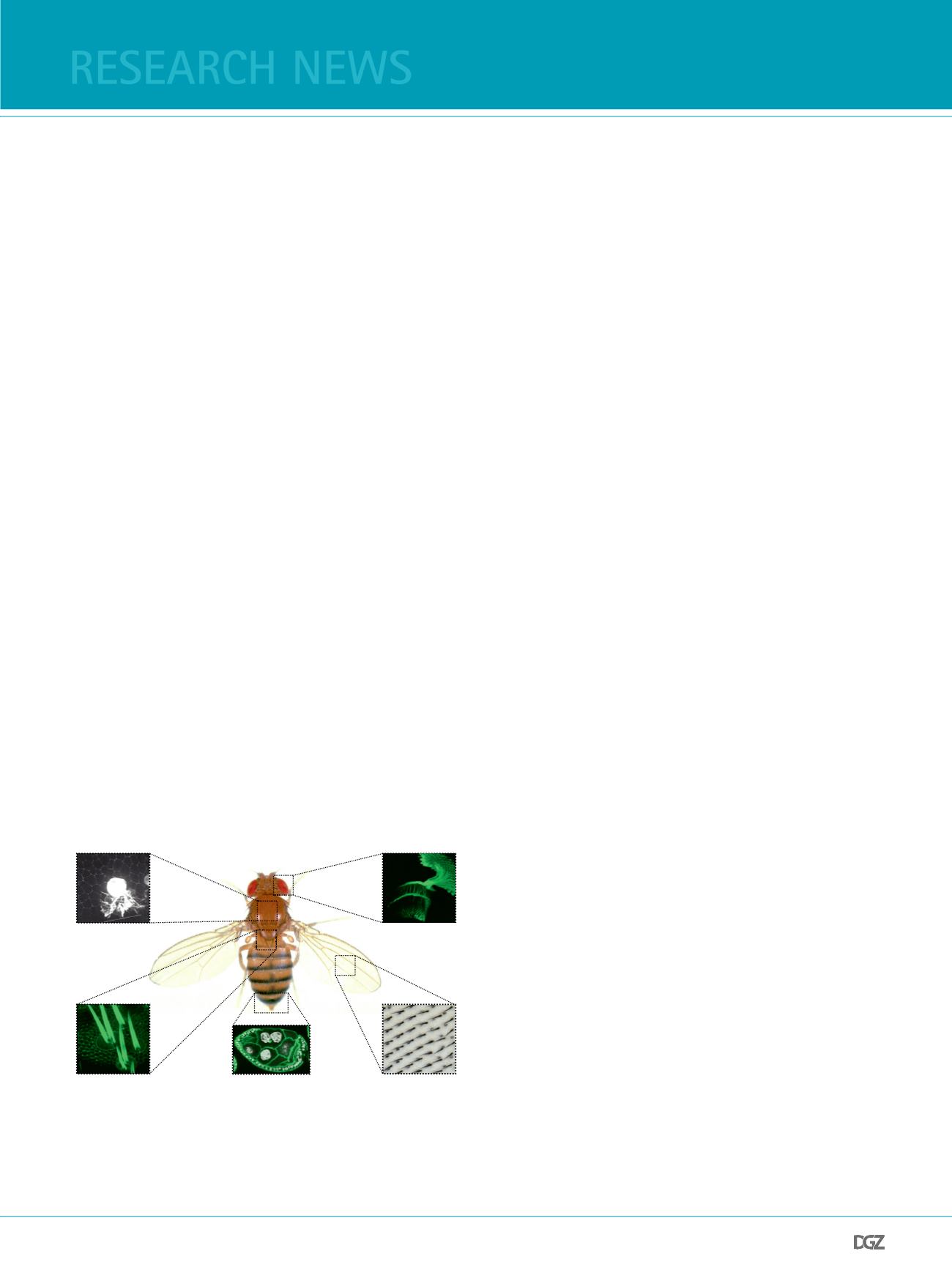
Epithelial polarization
of the wing epithelium
Hemocyte migration
and phagocytosis
in immune response
Development of
sensory organs
Axonal targeting in
the visual system
oogenesis
Figure 2: Actin driven processes in Drosophila development.
Despite their similar biochemical properties WASP proteins fulfll distinct
cellular functions in vivo during Drosophila development. For references
see text.


