Cell News 2/2014
14
Research news
Werner Risau Prize 2014
Coupling of angiogenesis and osteogenesis by a specific vessel
subtype in bone
Anjali P. Kusumbe
In the mammalian skeletal system, growth of the vascular net-
work is regulated by signals provided by chondrocytes and other
bone cells, among which the vascular endothelial growth factor
(VEGF) is studied best1. Conversely, blood vessels are thought to
influence the osteogenic generation of new bone2. In addition
to historic studies highlighting the close proximity of vascular
and osteoblastic cells, potential roles of angiogenic blood ves-
sel growth in fracture healing have been proposed3. It also has
been suggested that alterations in the skeletal microvasculature
might be linked to compromised hematopoiesis and osteogenesis
in human subjects with primary osteoporosis or at old age4,5.
However, direct evidence for such disease-causing or age-rela-
ted alterations is lacking and our understanding of the normal
organisation, functional specialization and precise function of
the skeletal vasculature is incomplete. The precise overall orga-
nization of the skeletal vasculature has remained poorly under-
stood because of technical difficulties associated with the pro-
cessing of bone combined with the loss of crucial 3D information
in thin tissue sections. Revised immunohistochemistry protocols
have now allowed us to image the bone vasculature at high re-
solution.
In addition to revised immunofluorescence protocols, we visua-
lised bone vessels with a combination of EC specific, tamoxifen-
inducible Cdh5(PAC)-CreERT2 and Rosa26-mT/mG Cre reporter
transgenic mice6,7. Imaging of the bone microvasculature with
both approaches uncovered structurally distinct capillary sub-
sets. Endothelial tubes in the metaphysis resembled straight co-
lumns that were interconnected by distal vessel loops or arches.
In contrast, diaphyseal capillaries displayed the highly branched
pattern characteristic for the sinusoidal vasculature of bone
marrow (Fig. 1a, b). At the interface between metaphysis and
diaphysis, the two vessel types were connected confirming that
they were part of one continuous vascular bed (Fig. 1b).
The different vessel types were distinguishable by immunos-
taining with specific cell surface markers. Columnar tubes and
arches in the metaphysis and endosteal endothelial cells (ECs)
were strongly positive for CD31/PECAM1 and Endomucin (Emcn),
while sinusoidal vessels in the diaphysis displayed only weak
CD31 staining and slightly lower Emcn expression (Fig. 1c). A
distinct CD31hi/Emcnhi endothelial subset could be also identi-
fied and separated from CD31lo/Emcnlo cells in single cell sus-
pensions of long bones(Fig. 1d). Quantitative analysis by flow
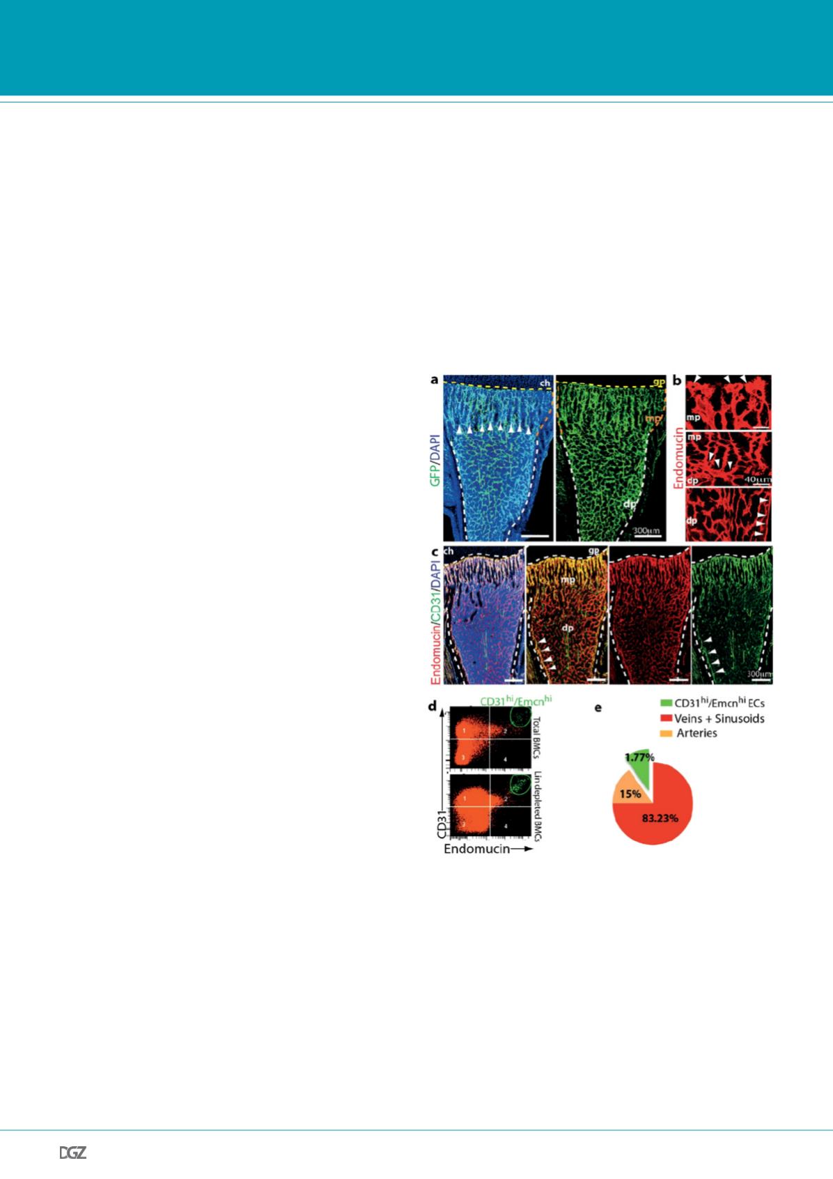
Figure 1. Identification of a distinct vessel subtype in murine bone.
a, Representative tile scan confocal images of the GFP+ (green) endotheli-
um in 4 week-old Cdh5(PAC)-CreERT2 x Rosa26-mT/mG double transgenic
tibia. Nuclei, DAPI (blue). Yellow dotted line marks growth plate (gp). Note
distinct organisation of microvessels in metaphysis (mp) and diaphysis (dp)
as well as their connections (arrowheads). b, Maximum intensity projec-
tions of Endomucin+ (red) column vessels in metaphysis (mp, arrowheads
mark distal protrusions) and highly branched sinusoids in diaphysis (dp).
Central panel shows interconnections (arrowheads) between both vessel
subtypes. c, Confocal tile scan of 4 week-old tibia images showing distinct
patterns of CD31+ (green) and Endomucin+ (red) ECs. Nuclei, DAPI (blue).
Strong CD31 and Endomucin signals mark capillaries in metaphysis (mp)
and endosteum (arrwoheads). d, Representative flow cytometry dot
plots showing the distinct CD31hi Endomucinhi EC subset in lineage (lin)
depleted bone marrow cells. e, Pie chart showing the relative abundance
of EC subtypes in 4 week-old long bone. CD31hi/Emcnhi cells represent
1.77±0.01% (mean ± s.d.m of 7 mice) of total ECs.


