Cell News 2/2014
15
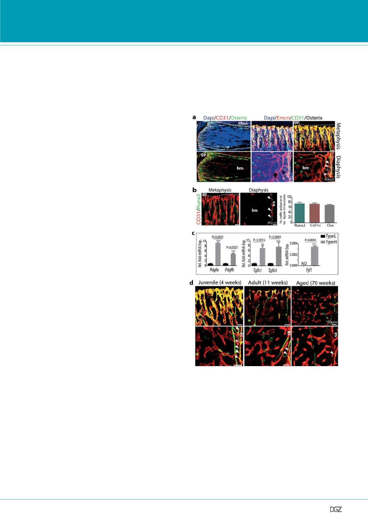
Figure 2. Association of osteoprogenitor cells with type H ECs and
decline of type H ECs in aged bones.
a. Confocal images of 4 week-old tibia with the indicated immunostai-
nings. Nuclei, DAPI (blue). Growth plate (gp) and bone marrow cavity (bm)
are marked. Osterix+ are found in proximity to CD31hi/Emcnhi (type H)
ECs in metaphysis and endosteum (arrowheads). b, Representative confocal
images (left panel) of immunostained 4 week-old tibia showing association
of Runx2+ osteoprogenitors (green) with CD31+ (red) vessels in metaphysis
and endosteum (es). Minimum exposure was used to capture CD31 fluore-
scence to project only cells with high CD31 intensity. Quantitative analysis
(right panel) of proximity (
≤
20µm) of Runx2+, Collagen1
α
+ (Col1
α
) and
Osterix+ (Osx) to nearest type H vessel. Mean±s.e.m, n=5. c, qPCR analysis
of growth factor expression (normalised to Actb) by CD31hi/Emcnhi ECs
relative to CD31lo/Emcnlo ECs sorted from murine tibia. Data represent
mean±s.e.m (n=4-6). P values, two-tailed unpaired t-test. d, Representa-
tive confocal images of CD31 (green) and Endomucin (red) immunostai-
ned tibia sections at 4, 11 and 70 weeks. Note age-dependent decline of
CD31hi/Endomucinhi ECs in metaphysis (upper panel) and in endosteal (es,
arrowheads) region in diaphysis (lower panel) of bone.
Research news
cytometry showed that CD31hi/Emcnhi cells represented only a
small fraction of total ECs (Fig. 1e). The observations above esta-
blished the existence of spatial and phenotypic heterogeneity in
the bone endothelium. On the basis of these findings, we propose
the following terminology for bone microvessels: type H for the
small CD31hi/Emcnhi subset and type L for the CD31lo/Emcnlo
sinusoidal vessels.
Immunostaining showed that Osterix+ osteoprogenitors, which
will give rise to osteoblasts and osteocytes8, were selectively po-
sitioned around type H but not type L endothelium (Fig. 2a). Des-
pite the low frequency (~1.77%) of type H endothelial cells (ECs)
in the bone endothelial cell fraction and ~0.015% in total bone
marrow (Fig. 1e), the majority of Runx2+ (82.63±1.8%), collagen
1
α
+ (74±3.3%) and Osterix+ cells (70±1.9%) were located di-
rectly adjacent to CD31hi/Emcnhi vessels (Fig. 2b,c).
To understand this distribution pattern of osteoblastic cells, the
expression of mRNAs for secreted growth factors with known
roles in osteoprogenitor survival and proliferation was analysed
in freshly purified ECs from long bone. Pdgfa, Pdgfb, Tgf1, Tgf3,
and Fgf1 transcripts were significantly higher expressed in type
H relative to type L ECs (Fig. 2c). Accordingly, the two bone ca-
pillary EC subsets have specific expression profiles suggesting
specialized functional properties.
It has been previously reported that osteoblast numbers declines
during aging9. Our analysis of bone endothelium during ageing
illustrated pronounced reduction of type H vessels, which were
much more abundant in juvenile (4 week-old) mice compared to
(11 week-old) adults, and were nearly absent in aged (70 week-
old) animals (Fig. 2d). EC proliferation was high within the type H
subpopulation in juvenile mice and declined rapidly in adulthood
(Data not shown). In contrast, the rate of type L EC proliferation
did not differ significantly between juvenile and older animals
(Data not shown).
HIF-1
α
controls physiological and pathological neo-angiogene-
sis2. To investigate HIF-1
α
function in the postnatal bone endo-
thelium, inducible EC-specific loss-of-function mice (Hif1ai
∆
EC)
were generated by combining loxP-flanked Hif1a alleles (Hi-
f1alox/lox)10 and Cdh5(PAC)-CreERT2 transgenics. Following
tamoxifen administration from postnatal day (P) 10 to P14,
analysis of Hif1ai
∆
EC mutants at P20 revealed striking vascular
defects. Type H endothelium was strongly reduced in metaphysis
and endosteum (Fig. 3a), the number of diaphyseal type L vessels
and ECs was comparable to control littermates.
The von Hippel-Lindau (VHL) E3 ubiquitin protein ligase controls
the stability and thereby biological activity of HIF-1
α
and other
substrates11. Inducible, EC-specific targeting of the murine Vhl
gene with the same strategy as described above for Hif1a led to
pronounced expansion of type H endothelium and metaphyseal
vessel columns and the surrounding osteoprogenitors (Fig. 3c,d).
Osteoprogenitors were significantly reduced in Hif1ai
∆
EC sam-
ples (Fig. 3a,b).


