Cell News 2/2014
16
Research news
Prolyl-4-hydroxylases (PHDs) modify HIF-1
α
and thereby mark
the protein for degradation under normoxic conditions. Accor-
dingly, PHD inhibitors, such as deferoxamine mesylate (DFM),
enhance HIF-1
α
stability and activity12. Next, we tested whe-
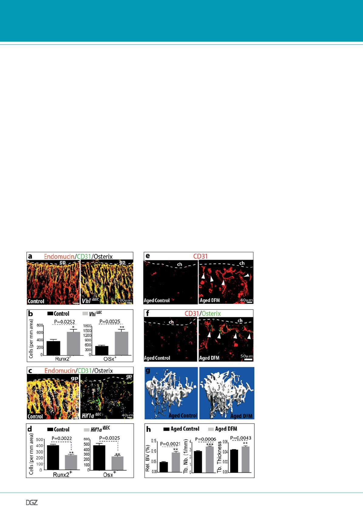
ther DFM promotes CD31hi/Emcnhi ECs, neo-angiogenesis and
osteogenesis in aged animals. While long bones of aged, 64 to
70 week-old mice treated with vehicle control contained very
few CD31hi/Emcnhi vessels, DFM administration led to substan-
tial expansion of type H endothelium (Fig. 3e) and emergence
of vessel-associated Osterix+ cells (Fig. 3f). Furthermore, µ-CT
examination showed that 6 weeks of DFM treatment led to sig-
nificantly increased bone mass (Fig. 3g,h). While the activity of
DFM is not restricted to ECs and is likely to affect multiple cell
populations, the findings above argue for crucial roles of endo-
thelial HIF in controlling bone angiogenesis, type H vessel abun-
dance, endothelial growth factor expression, and osteogenesis.
Above finding that capillaries in the skeletal system of mice can
be subdivided into type H and type L endothelium on the ba-
sis of morphological, molecular and functional criteria should
be hugely beneficial for future studies in basic and medical re-
search. CD31hi/Emcnhi capillaries at the distal end of the arterial
network in bone might represent the central building block of
a metabolically specialised tissue environment with privileged
access to oxygen and nutrients, which is likely to influence the
growth potential and metabolism of other cell types. This is not
only relevant for osteoblastic cells but potentially also for hema-
topoietic stem and progenitor cells, which preferentially home to
the metaphysis after transplantation13.
We also propose that type H ECs mediate local growth of the
vasculature and provide niche signals for perivascular osteopro-
genitors. Type H vessel formation and the expression of potential
angiocrine factors for osteoblastic cells are enhanced by HIF and
by Notch signalling14. Thus, the abundance of CD31hi/Emcnhi
ECs may be useful as diagnostic readout for the growth status of
the bone vasculature and its pro-osteogenic capacity. Our results
also indicate that specific molecular pathways can be used to
boost type H vessel formation and osteogenesis. This might be
of great importance for conditions involving compromised frac-
ture healing or loss of bone mass. Ageing and post-menopausal
estrogen deficiency are major risk factors for osteoporosis, and
estrogen can promote angiogenesis15. Accordingly, decline of
type H vessels and the concomitant reduction of osteoprogeni-
tor cells could potentially offer a compelling explanation for the
loss of bone mass during ageing and might enable therapeutic
Figure 3. Type H ECs couple angio-
genesis and osteogenesis.
a, Representative confocal images
of CD31 (green), Endomucin (red)
and Osterix (white) immunos-
tained, 3 week-old Vhli
∆
EC and
control tibiae. b, Quantitation of
Runx2+ and Osterix+ in Vhli
∆
EC
mutants and littermate controls.
Data represent mean±s.e.m (n=5). P
values, two-tailed unpaired t-test.
c, Maximum intensity projections of
3 week-old Hif1ai
∆
EC and con-
trol tibia stained for CD31 (green),
Endomucin (red) and Osterix (white).
Growth plate, gp. d, Quantitation
of Runx2+ and Osterix+ cells in
Hif1ai
∆
EC mutant and control long
bone. Data represent mean±s.e.m
(n=5). P values, two-tailed unpaired
t-test. e, f, Representative confocal
images of CD31 (red, f) or CD31
and Osterix (green, g) stained tibia
sections from aged DFM-treated and
control mice. Low intensity projec-
tion shows only CD31hi cells. DFM
induces CD31hi vessels and Osterix+
osteoprogenitors. Chondrocytes, ch.
g, Representative µ-CT images of
tibias from aged DFM-treated and
control mice. h, Quantitative µ-CT
analysis of relative bone volume (Rel.
BV), trabecular number (Tb. Nb), and
trabecular thickness (Tb. Thickness) in
proximal tibia from aged DFM-trea-
ted and control mice. Data represent
mean±s.e.m (n=5). P values, two-
tailed unpaired t-test.


